. 2002 Feb;13(2):607-20.
doi: 10.1091/mbc.01-08-0400.

Drosophila minichromosome maintenance 6 is required for chorion gene amplification and genomic replication

Affiliations

Drosophila minichromosome maintenance 6 is required for chorion gene amplification and genomic replication

Gina Schwed et al. Mol Biol Cell. 2002 Feb.

Abstract

Duplication of the eukaryotic genome initiates from multiple origins of DNA replication whose activity is coordinated with the cell cycle. We have been studying the origins of DNA replication that control amplification of eggshell (chorion) genes during Drosophila oogenesis. Mutation of genes required for amplification results in a thin eggshell phenotype, allowing a genetic dissection of origin regulation. Herein, we show that one mutation corresponds to a subunit of the minichromosome maintenance (MCM) complex of proteins, MCM6. The binding of the MCM complex to origins in G1 as part of a prereplicative complex is critical for the cell cycle regulation of origin licensing. We find that MCM6 associates with other MCM subunits during amplification. These results suggest that chorion origins are bound by an amplification complex that contains MCM proteins and therefore resembles the prereplicative complex. Lethal alleles of MCM6 reveal it is essential for mitotic cycles and endocycles, and suggest that its function is mediated by ATP. We discuss the implications of these findings for the role of MCMs in the coordination of DNA replication during the cell cycle.


Figures
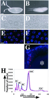
Figure 1
Figure 1
The eggshell and cellular phenotype of fs(1)K1214. (A) An egg from a wild-type mother is turgid with a thick eggshell that includes two prominent, rigid dorsal appendages. (B) An egg from a homozygous fs(1)K1214 mother is flaccid with a thin eggshell and flimsy dorsal appendages. Anterior is to the left and dorsal is up. Bar, 100 μm (A and B). (C) A 40× phase contrast image of a wild-type eggshell has a hexagonal pattern. Each hexagon represents a “footprint” of the follicle cell that formed that unit of the eggshell late in oogenesis before it died and was sloughed off. (D) Dorsal view of an eggshell produced by an fs(1)K1214 mother shows that it is less phase dense than wild-type, although some follicle cell footprints are evident in the dorsal/anterior on the left. Bar (C and D), 10 μm. (E) BrdU labeling (red) in wild-type stage 10B follicle cell nuclei (blue) reveals four spots of incorporation. The two most prominent spots are the amplifying chorion loci on the X and 3rd chromosome, whereas the faint spots represent unknown loci. (F) Most mutant fs(1)K1214 follicle cells in stage 10B have undetectable BrdU incorporation, whereas a few have faint or nearly wild-type incorporation at amplifying loci. Images in E and F represent a composite stack of eight, 1-μm confocal sections. Bar, 10 μm. (G) Lateral confocal image of a stage 12 egg chamber in an fs(1)K1214 ovary. Shown are the dorsal anterior follicle cells that are mostclosely apposed to the oocyte nucleus (asterisk), and which have the most robust BrdU labeling at chorion loci and elsewhere in the nucleus. Image represents a composite stack of 16, 1 μm confocal sections. Dorsal is up and anterior is to the left. (H) Flow sorting of DAPI stained nuclei from wild-type (blue) and fs(1)K1214 (magenta) ovaries indicates there is no significant difference in DNA content between them. The smaller peaks from the less abundant, but higher ploidy, nurse cells also gave no evidence for endocycle defects in fs(1)K1214 (our unpublished results).
Figure 2
Figure 2
Genetic and molecular mapping of fs(1)K1214. A molecular map showing the position of the MCM6 transcription unit (black box) relative to the two P elements (triangles) used to generate new deficiencies. The new deficiencies are indicated below. Solid lines represent deleted regions and dotted lines represent uncertainty in the extent of the deficiencies. Arrows indicate that the deficiency extends beyond the region shown (see MATERIALS AND METHODS). Whether the deficiencies complemented the fs(1)K1214 thin eggshell phenotype is indicated on the right. The two deficiencies that failed to complement deleted MCM6, whereas those that complemented did not delete the gene. Not shown are two lethal excision strains (6C-166 and 6C-157) that complemented, and in which we did not detect a deletion.
Figure 3
Figure 3
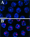
MCM6 rescues fs(1)K1214. (A and B) BrdU labeling (red) in stage 10B follicle cell nuclei (blue) from females homozygous for fs(1)K1214 without (A) and with (B) 1 copy of Ub:FL:MCM6. Images represent a composite stack of eight, 1-μm confocal sections. Bar, 10 μm.
Figure 4
Figure 4
MCM6 associates with MCM2, 4, and 5 during amplification. Western blot of immunoprecipitation with anti-FLAG antibody by using extracts from ovaries of c323GAL4; UAS:FL:MCM6 (UFM), which expresses FLAG:MCM6 specifically in follicle cells during amplification, or the transformation host lacking the transgene (y w). I = 1/500 of input from total extract, p = 1/10 pellet. Duplicate blots were probed with antibodies against FLAG, MCM2, MCM4, and MCM5 giving evidence for coimmunoprecipitation of FLAG:MCM6 with MCM2 and 4, and, minimally, with MCM5. Note that the input for MCM2, 4, and 5 is from all cells in the ovary, whereas the pellet represents protein precipitated with FLAG:MCM6 expressed only in amplifying follicle cells. Equal amounts of y w and UFM were loaded based on measurement of total protein in the input extract.
Figure 5
Figure 5
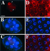
MCM6 is chromatin associated but not visibly concentrated at chorion loci. (A-C) Stage 4 egg chamber from the Ub:FL:MCM6 strain stained with DAPI (B) and anti-FLAG antibody (A). DAPI staining in B shows four of the 15 nurse cells in the central part of the egg chamber that have condensed polytene chromosomes at this stage. The smaller nuclei on the periphery are within follicle cells. Anti-FLAG staining in A and merged image in C shows that MCM6 is associated with chromosomes in only two of these nuclei (arrowheads) consistent with asynchronous DNA replication in these egg chambers. (D and F) Anti-FLAG staining of stage 10B follicle cells from the UAS:FL:MCM6 strain does not reveal focal staining corresponding to chorion loci. (D) Anti-FLAG (E) DAPI staining (F) merged image. The intense DAPI spots in E correspond to the heterochromatic chromocenter. Bar, 10 μm.
Figure 6
Figure 6
MCM6 mutant brains have a severe reduction in DNA replication and cell proliferation. (A) Brain hemisphere from a wild-type late 3rd instar larva labeled with BrdU (red) to detect proliferating cells in S phase. Labeling reveals numerous cells in S phase in the outer proliferation center (OPC), inner proliferation center (IPC), and midbrain (MB). (B) Brain from an MCM6 mutant larva of the same stage is greatly reduced in size and has few cells that detectably incorporate BrdU. A and B are the same magnification and exposure. Bar (A and B), 100 μm. (C) Cluster of cells from the midbrain of a wild-type larva that are in S phase has robust BrdU incorporation in the nucleus. These represent the nuclei of large neuroblast stem cells (NB) and its smaller descendants. (D) Nuclei from MCM6 mutant cells have greatly reduced incorporation of BrdU into small foci. The larger foci (one indicated by arrowhead) represent heterochromatin that replicates late in S phase. Bar (C and D), 10 μm.
Figure 7
Figure 7
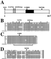
Sequence of MCM6 mutant alleles. (A) Amino acid changes in the MCM6 mutant alleles are indicated above a schematic representation of the protein. The fs(1)K1214 mutation is homozygous viable and changes a methionine at 676 to a lysine (M676K). The four other mutations are homozygous lethal. The black shaded portion represents the highly conserved MCM box. Gray shading indicates the putative C4 Zinc finger. See Table 1 for allele numbers. M, methionine; I, isoleucine; T, threonine; Q, glutamine; K, lysine. (B-D) ClustalW alignment of selected regions of Drosophila MCM6 protein with MCM6 proteins from other species is shown to indicate the degree of conservation of the amino acid residues that are changed in the mutants (arrows above). (B) Lethal T157M mutation changes a highly conserved threonine that lies between the cysteine pairs (asterisks above) of a putative C4 Zinc finger motif. The Q165stop mutation predicts a translation stop within this motif. (C) Lethal G388D mutation lies within a sequence similar to the Walker A motif (bracket below) that is conserved among proteins that bind and hydrolyze ATP. (D) Viable but amplification defective M676K mutation lies within a region of low conservation between Drosophila and other MCM6 proteins. Not shown is M1I, which mutates the putative initiator methionine. Conserved regions are boxed. Dark shading indicates identical residues. Light shading indicates conservative substitutions. No shading indicates residues that are not conserved. D.m., Drosophila melanogaster; H.s., Homo sapiens; M.m., Mus musculus; X.l., Xenopus laevis; S.p., Schizosaccharomyces pombe; S.c., S. cerevisiae.
Figure 8
Figure 8
Schematic model for the composition and regulation of the Amplification Complex (AC). Based on our results and those of previous investigations, it is likely that an AC resembling the pre-RC assembles onto chorion origins. Current evidence suggests that the AC contains most of the components that comprise pre-RCs at other origins (colored ovals), and responds to S-phase kinases (boxes). CDC6 (white oval) is the only known pre-RC component that has not been linked to amplification. Unlike the pre-RC, the AC may contain one or more amplification factors (AF) (black pentagon) that permit rereplication in the presence of constitutively high CDK activity. Recent evidence suggests that E2F1 and RBF1 participate directly in the regulation of chorion and other origins. See text for references.

Similar articles

Cited by

References

    1. Adams MD, et al. The genome sequence of Drosophila melanogaster. Science. 2000;287:2185–2195. - PubMed
    1. Aparicio O, Weinstein D, Bell S. Components and dynamics of DNA replication complexes in S. cerevisiae: redistribution of MCM proteins and Cdc45p during S phase. Cell. 1997;91:59–69. - PubMed
    1. Asano M, Wharton RP. E2F mediates developmental and cell cycle regulation of ORC1 in Drosophila. EMBO J. 1999;18:2435–2448. - PMC - PubMed
    1. Austin RJ, Orr-Weaver TL, Bell SP. Drosophila ORC specifically binds to ACE3, an origin of DNA replication control element. Genes Dev. 1999;13:2639–2649. - PMC - PubMed
    1. Bell S, Stillman B. ATP-dependent recognition of eukaryotic origins of DNA replication by a multiprotein complex. Nature. 1992;357:128–134. - PubMed

Publication types

Substances