. 2002 Feb;13(2):656-69.
doi: 10.1091/mbc.01-09-0454.

Dictyostelium LvsB mutants model the lysosomal defects associated with Chediak-Higashi syndrome

Affiliations

Dictyostelium LvsB mutants model the lysosomal defects associated with Chediak-Higashi syndrome

Edward Harris et al. Mol Biol Cell. 2002 Feb.

Abstract

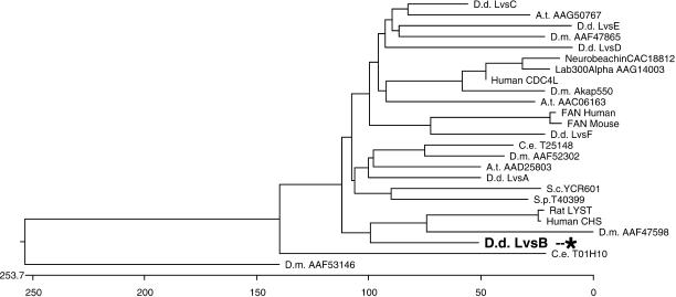
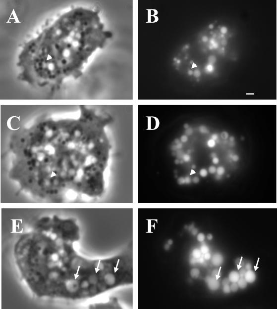
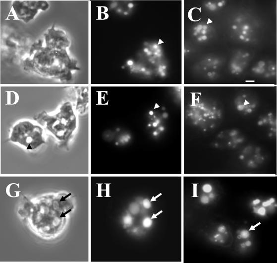
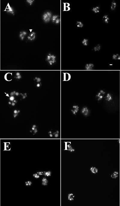
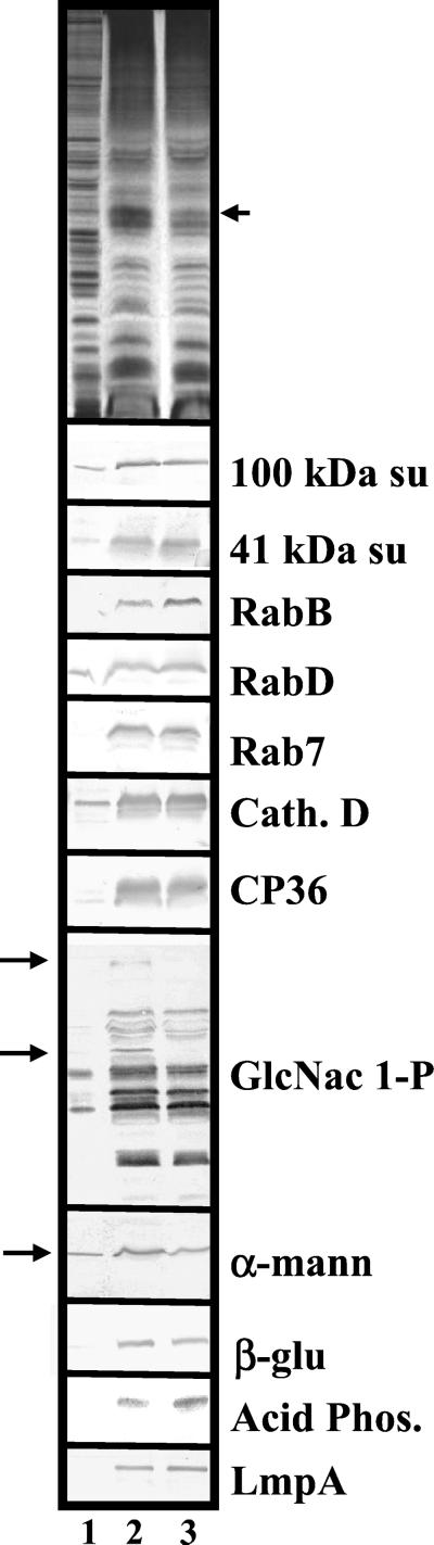
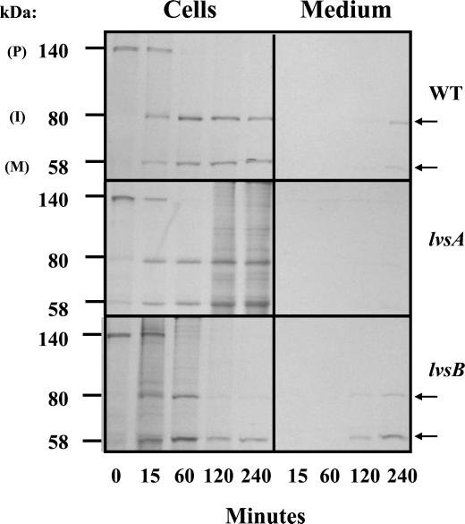
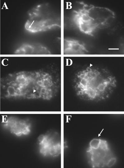
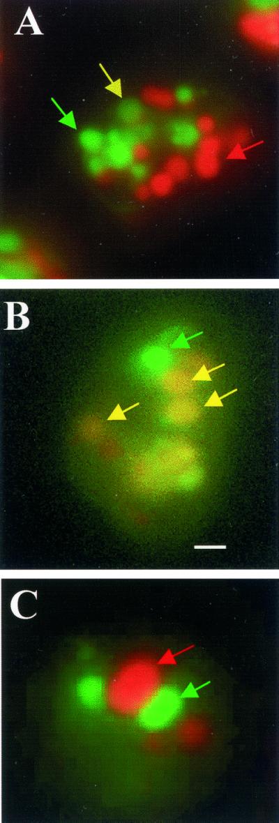
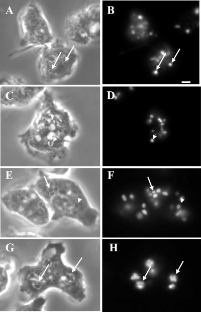
Chediak-Higashi syndrome is a genetic disorder caused by mutations in a gene encoding a protein named LYST in humans ("lysosomal trafficking regulator") or Beige in mice. A prominent feature of this disease is the accumulation of enlarged lysosome-related granules in a variety of cells. The genome of Dictyostelium discoideum contains six genes encoding proteins that are related to LYST/Beige in amino acid sequence, and disruption of one of these genes, lvsA (large volume sphere), results in profound defects in cytokinesis. To better understand the function of this family of proteins in membrane trafficking, we have analyzed mutants disrupted in lvsA, lvsB, lvsC, lvsD, lvsE, and lvsF. Of all these, only lvsA and lvsB mutants displayed interesting phenotypes in our assays. lvsA-null cells exhibited defects in phagocytosis and contained abnormal looking contractile vacuole membranes. Loss of LvsB, the Dictyostelium protein most similar to LYST/Beige, resulted in the formation of enlarged vesicles that by multiple criteria appeared to be acidic lysosomes. The rates of endocytosis, phagocytosis, and fluid phase exocytosis were normal in lvsB-null cells. Also, the rates of processing and the efficiency of targeting of lysosomal alpha-mannosidase were normal, although lvsB mutants inefficiently retained alpha-mannosidase, as well as two other lysosomal cysteine proteinases. Finally, results of pulse-chase experiments indicated that an increase in fusion rates accounted for the enlarged lysosomes in lvsB-null cells, suggesting that LvsB acts as a negative regulator of fusion. Our results support the notion that LvsB/LYST/Beige function in a similar manner to regulate lysosome biogenesis.


Figures

Figure 1
Figure 1
LvsB is the Dictyostelium protein most similar to mammalian LYST/Beige. Sequence comparison of the BEACH and WD domains of the six Dictyostelium Lvs proteins and proteins from other organisms. The BEACH and WD domains of the indicated proteins were aligned using DNAStar with the ClustalW algorithm. The position of LvsB is indicated with an asterisk. Note its close relationship to mammalian LYST proteins. Accession codes are indicated for each protein. A.t., Arabidopsis thaliana; C.e., Caenorhabditis elegans; D.d., Dictyostelium discoideum; D.m., Drosophila melanogaster.
Figure 2
Figure 2
The enlarged vesicles in the lvsB-null mutant are endocytic. NC4A2 control (A and B), lvsA-null (C and D), and lvsB-null (E and F) cells were incubated in HL-5 medium containing FITC-dextran for 1 h to illuminate the endocytic vesicles. Cells were visualized using a fluorescence microscope. The arrows point to enlarged endocytic vesicles in lvsB-null cells, and the arrowheads point to vesicles the approximate size of normal lysosomes. Bar, 1 μm.
Figure 3
Figure 3
The enlarged vesicles in the lvsB-null cells are acidic lysosomes. To visualize lysosomes, NC4A2 control (A and B), lvsA-null (D and E), and lvsB-null (G and H) cells were subjected to a 15-min pulse with FITC-dextran, washed, and chased for 15 min in fresh growth medium. Cells were examined with phase-contrast optics (A, D, and G) or by fluorescence microscopy (B, E, and H). The control (B) and lvsA-null (E) cells contained lysosomes of normal size, whereas the lvsB-null cells (H) contained enlarged lysosomes. Control (C), lvsA-null (F), and lvsB-null (I) cells were incubated with the acidophilic dye LysoSensor DND-189 (Molecular Probes) in HL-5 growth medium and visualized using a fluorescence microscope. The arrows point to enlarged acidic lysosomes, and the arrowheads point to normal size lysosomes. Bar, 2 μm.
Figure 4
Figure 4
Only the lvsB-null cells exhibit the CHS cellular morphology. To examine the size of the endocytic vesicles, control (A), lvsA-null (B), lvsB-null (C), lvsC-null (D), lvsD-null (E), and lvsE-null (F) cells were pulsed in HL-5 medium with FITC-dextran for 2 h, washed, and fixed (lvsF-null not shown). Vesicle morphology was examined with a fluorescent microscope at 400× magnification. Bar, 2 μm.
Figure 5
Figure 5
Lysosomes from the lvsB-null mutant reduced levels of α-mannosidase and several cysteine proteinases. NC4A2 control and lvsB-null cells were fed iron-dextran for 15 min and then were chased for 15 min in fresh medium to load up lysosomes. Iron-dextran–filled vesicles (lysosomes) were collected on a magnetic column, concentrated, and subjected to SDS-PAGE (see MATERIALS AND METHODS). Control cell lysates (lane 1), control lysosomes (lane 2), and lvsB-null lysosomes (lane 3) were subjected to SDS-PAGE and silver stained (top) or incubated with a host of antibodies against lysosomal proteins. The antibodies reacted with the 100-kDa subunit of the vacuolar H+-ATPase (100 kDa su), the 41-kDa subunit of the vacuolar H+-ATPase (41 kDa su), RabB (human Rab21 homolog), RabD (human Rab14 homolog), Rab7, cathepsin D (Cath. D), cysteine proteinase 36 (CP36), GlcNac 1-P-sulfate (lysosomal cysteine proteinases), α-mannosidase (α-mann), β-glucosidase (β-glu), acid phosphatase (Acid Phos.), and lysosomal membrane protein A (LmpA).
Figure 6
Figure 6
lvsB-null cells process and target lysosomal α-mannosidase normally but oversecrete the mature enzyme. Wild-type control, lvsA-null, and lvsB-null cells were pulsed with [35S]methionine, washed, and chased in fresh HL-5. At each time point during the chase period, α-mannosidase was immunoprecipitated from both the medium, to detect secreted α-mannosidase, and cell lysates, to detect intracellular α-mannosidase. Immunoprecipitates were subjected to SDS-PAGE, followed by fluorography.
Figure 7
Figure 7
The rates of endocytosis, phagocytosis, and fluid exocytosis are comparable between lvsB-null and control cells; lvsA-null cells are defective in phagocytosis. Endocytosis rates (A), exocytosis rates (B), and phagocytosis rates (C) were measured for control cells and the lvsA-null and lvsB-null mutants as described in MATERIALS AND METHODS. lvsA-null cells were significantly reduced in the rate of phagocytosis (unpaired, two-tailed Student's t test, p < 0.01) compared with control cells, whereas, lvsB-null and control cells were not significantly different.
Figure 8
Figure 8
The CV system of membranes appears normal in the lvsB-null cells in contrast to the altered morphology seen for CV membranes in lvsA-null cells. Control NC4A2 (A and B), lvsA-null (C and D), and lvsB-null (E and F) were fixed, permeabilized, and incubated with an antibody against the 100-kDa subunit of the proton pump, a marker for the CV membranes. CV reticular network membranes were visualized with a fluorescent microscope at 1000×.
Figure 9
Figure 9
Large endolysosomes in the lvsB-null cells are a result of an increase in fusion. Control NC4A2, lvsA-null, and lvsB-null cells were pulsed with RITC-dextran, washed, pulsed with FITC-dextran, washed, chased for 5 min, fixed, and examined under a fluorescence microscope. Most of the red and green vesicles were distinct and separate from each other in the control (A), whereas, a higher percentage of vesicles in the lvsB-null (B) fused with each other. Examination of cells at the end of the double-pulse period revealed that little colocalization was observed in the mutant, suggesting that separately internalized vesicles fuse over time (C). Bar, 2 μm.
Figure 10
Figure 10
The rate of phagosome-phagosome fusion was increased in lvsB-null cells. Control cells were pulsed with FITC-labeled Escherichia coli for 10 min, washed, and chased on coverslips for 30 min. At this time point indicated, cells were fixed and examined microscopically to determine the number of fusion events. The assay is linear between 30 and 90 min of chase. A, C, E, and G are phase contrast images, whereas B, D, F, and H are fluorescent images. A and B (control) and C and D (mutant) indicate that after a short pulse period only single-particle–containing phagosomes are observed in cells (marked with arrowheads). E and F (control) and G and H (mutant) indicate that at the 30-min chase point at greater number of phagosomes contain multiparticles in the mutant cells as compared with control cells (marked with arrows). Bar, 2 μm.
Figure 11
Figure 11
The rate of endolysosomal and phagosomal fusion was increased in lvsB-null cells. A enumerates the number of red/green-merged endosomal vesicles and reveals that this number is significantly higher in the lvsB-null cells relative to control (wt) and lvsA-null cells (unpaired, two-tailed Student's t test, p = 0.0016). B illustrates that the number of fusion events per cell is significantly higher at the 30-min chase point in the lvsB-null mutant as compared with lvsA-null and control cells (wt) when using FITC-labeled bacteria (see MATERIALS AND METHODS for details) as a marker for phagolysosomes (unpaired, two-tailed Student's t test, p < 0.0001). These experiments were repeated three times each and for each experiment between 50 and 100 cells in random fields were analyzed.

Similar articles

Cited by

References

    1. Adam-Klages S, Adam D, Wiegmann K, Struve S, Kolanus W, Schneider-Mergener J, Kronke M. FAN, a novel WD-repeat protein, couples the p55 TNF-receptor to neutral sphingomyelinase. Cell. 1996;86:937–947. - PubMed
    1. Bahadoran P, Aberdam E, Mantoux F, Busca R, Bille K, Yalman N, de Saint-Basile G, Casaroli-Marano R, Ortonne JP, Ballotti R. Rab27a: a key to melanosome transport in human melanocytes. J Cell Biol. 2001;152:843–850. - PMC - PubMed
    1. Barrat FJ, Le Deist F, Benkerrou M, Bousso P, Feldmann J, Fischer A, de Saint B. Defective CTLA-4 cycling pathway in Chediak-Higashi syndrome: a possible mechanism for deregulation of T lymphocyte activation. Proc Natl Acad Sci USA. 1999;96:8645–8650. - PMC - PubMed
    1. Bucci C, Thomsen P, Nicoziani P, McCarthy J, van Deurs B. Rab7: a key to lysosome biogenesis. Mol Biol Cell. 2000;11:467–480. - PMC - PubMed
    1. Buczynski G, Bush J, Zhang L, Rodriguez-Paris J, Cardelli J. Evidence for a recycling role for Rab7 in regulating a late step in endocytosis and in retention of lysosomal enzymes in Dictyostelium discoideum. Mol Biol Cell. 1997;8:1343–1360. - PMC - PubMed

Publication types

LinkOut - more resources