

The interaction between the coactivator dCBP and Modulo, a chromatin-associated factor, affects segmentation and melanotic tumor formation in Drosophila
- PMID: 11854460
- PMCID: PMC122444
- DOI: 10.1073/pnas.052509799
The interaction between the coactivator dCBP and Modulo, a chromatin-associated factor, affects segmentation and melanotic tumor formation in Drosophila
Abstract
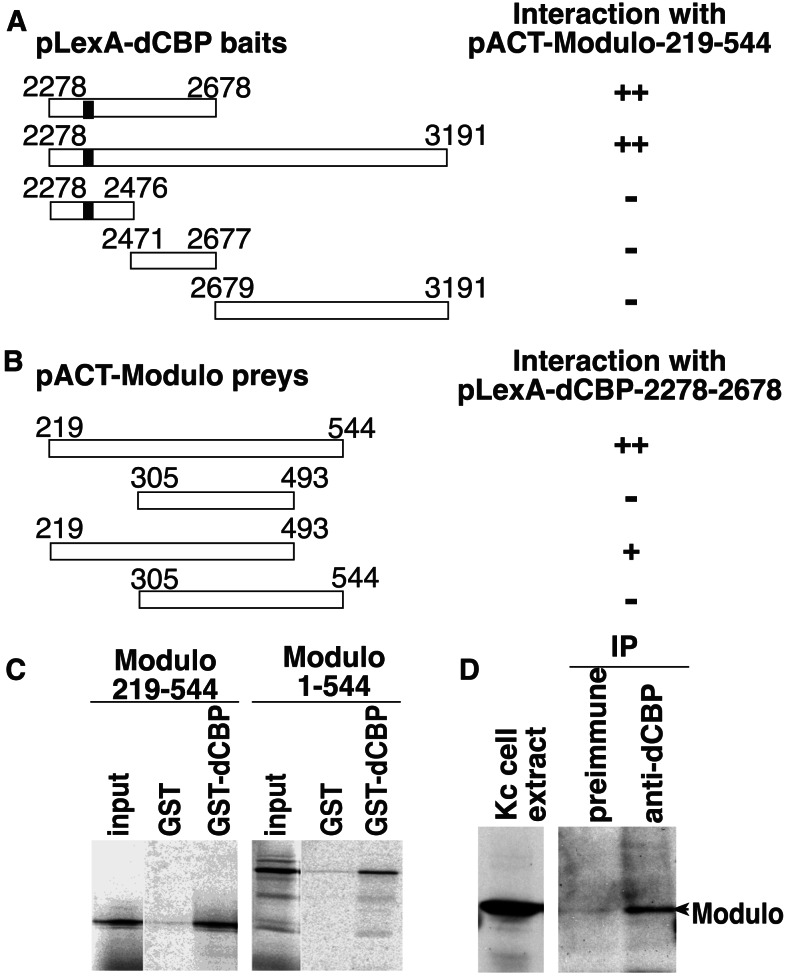
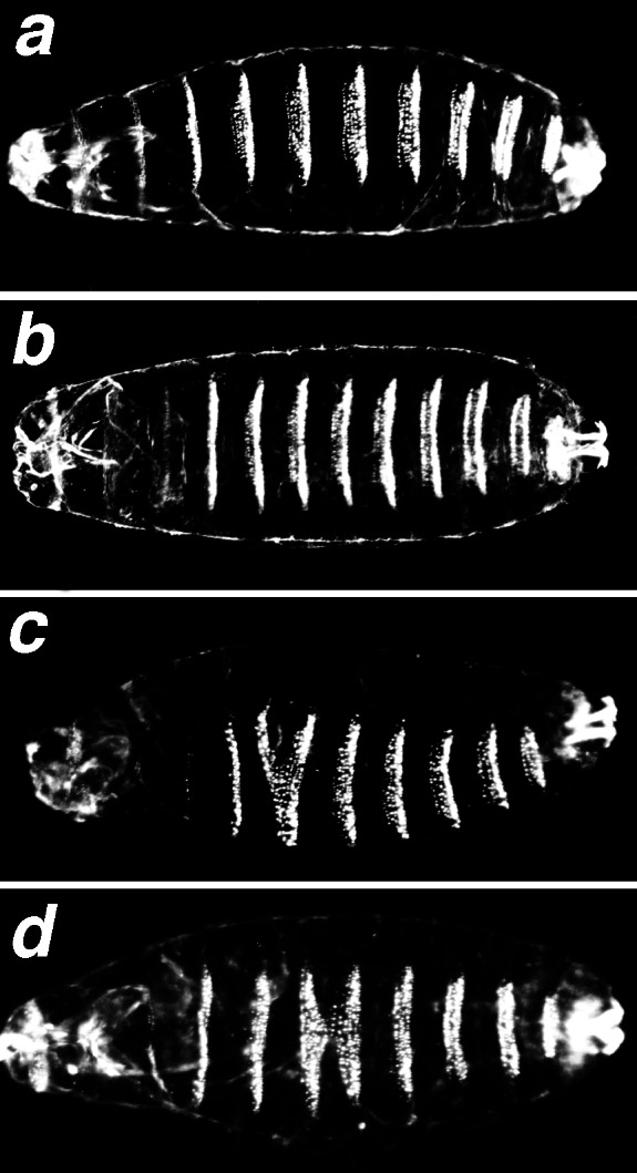
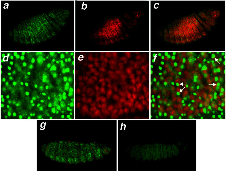
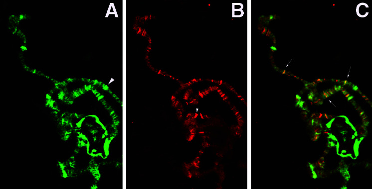
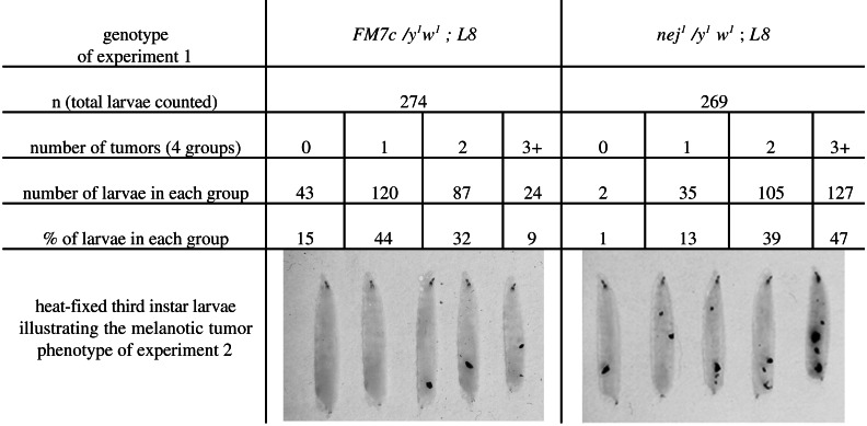
The development of Drosophila requires the function of the CREB-binding protein, dCBP. In flies, dCBP serves as a coactivator for the transcription factors Cubitus interruptus, Dorsal, and Mad, and as a cosuppressor of Drosophila T cell factor. Current models propose that CBP, through its intrinsic and associated histone acetyltransferase activities, affects transient chromatin changes that allow the preinitiation complex to access the promoter. In this report, we provide evidence that dCBP may regulate the formation of chromatin states through interactions with the modulo (mod) gene product, a protein that is thought to be involved in chromatin packaging. We demonstrate that dCBP and Modulo bind in vitro and in vivo, that mutations in mod enhance the embryonic phenotype of a dCBP mutation, and that dCBP mutations enhance the melanotic tumor phenotype characteristic of mod homozygous mutants. These results imply that, in addition to its histone acetyltransferase activity, dCBP may affect higher-order chromatin structure.
Figures
Similar articles
-
Functional interaction between the coactivator Drosophila CREB-binding protein and ASH1, a member of the trithorax group of chromatin modifiers.Mol Cell Biol. 2000 Dec;20(24):9317-30. doi: 10.1128/MCB.20.24.9317-9330.2000. Mol Cell Biol. 2000. PMID: 11094082 Free PMC article.
-
Cubitus interruptus requires Drosophila CREB-binding protein to activate wingless expression in the Drosophila embryo.Mol Cell Biol. 2000 Mar;20(5):1616-25. doi: 10.1128/MCB.20.5.1616-1625.2000. Mol Cell Biol. 2000. PMID: 10669739 Free PMC article.
-
The acetyltransferase activity of CBP is required for wingless activation and H4 acetylation in Drosophila melanogaster.Mol Cell Biol. 2002 Jun;22(11):3832-41. doi: 10.1128/MCB.22.11.3832-3841.2002. Mol Cell Biol. 2002. PMID: 11997517 Free PMC article.
-
Chromatin mechanisms in Drosophila dosage compensation.Prog Mol Subcell Biol. 2005;38:123-49. doi: 10.1007/3-540-27310-7_5. Prog Mol Subcell Biol. 2005. PMID: 15881893 Review.
-
CBP, a transcriptional coactivator and acetyltransferase.Biochem Cell Biol. 2001;79(3):253-66. Biochem Cell Biol. 2001. PMID: 11467739 Review.
Cited by
-
Biological functions of the ISWI chromatin remodeling complex NURF.Genes Dev. 2002 Dec 15;16(24):3186-98. doi: 10.1101/gad.1032202. Genes Dev. 2002. PMID: 12502740 Free PMC article.
-
Analysis of Drosophila melanogaster proteome dynamics during embryonic development by a combination of label-free proteomics approaches.Proteomics. 2016 Aug;16(15-16):2068-80. doi: 10.1002/pmic.201500482. Epub 2016 May 10. Proteomics. 2016. PMID: 27029218 Free PMC article.
-
Zika virus infection drives epigenetic modulation of immunity by the histone acetyltransferase CBP of Aedes aegypti.PLoS Negl Trop Dis. 2022 Jun 27;16(6):e0010559. doi: 10.1371/journal.pntd.0010559. eCollection 2022 Jun. PLoS Negl Trop Dis. 2022. PMID: 35759510 Free PMC article.
-
Corepressive action of CBP on androgen receptor transactivation in pericentric heterochromatin in a Drosophila experimental model system.Mol Cell Biol. 2009 Feb;29(4):1017-34. doi: 10.1128/MCB.02123-07. Epub 2008 Dec 15. Mol Cell Biol. 2009. PMID: 19075001 Free PMC article.
-
Transcriptional regulation by Modulo integrates meiosis and spermatid differentiation in male germ line.Proc Natl Acad Sci U S A. 2006 Aug 8;103(32):11975-80. doi: 10.1073/pnas.0605087103. Epub 2006 Jul 28. Proc Natl Acad Sci U S A. 2006. PMID: 16877538 Free PMC article.
References
Publication types
MeSH terms
Substances
LinkOut - more resources
Full Text Sources
Molecular Biology Databases
