. 2002 Feb 19;99(4):1960-5.
doi: 10.1073/pnas.022638399.

Mechanical disruption of individual nucleosomes reveals a reversible multistage release of DNA

Affiliations

Mechanical disruption of individual nucleosomes reveals a reversible multistage release of DNA

Brent D Brower-Toland et al. Proc Natl Acad Sci U S A. .

Abstract

The dynamic structure of individual nucleosomes was examined by stretching nucleosomal arrays with a feedback-enhanced optical trap. Forced disassembly of each nucleosome occurred in three stages. Analysis of the data using a simple worm-like chain model yields 76 bp of DNA released from the histone core at low stretching force. Subsequently, 80 bp are released at higher forces in two stages: full extension of DNA with histones bound, followed by detachment of histones. When arrays were relaxed before the dissociated state was reached, nucleosomes were able to reassemble and to repeat the disassembly process. The kinetic parameters for nucleosome disassembly also have been determined.


Figures

Figure 1
Figure 1
Disruption of individual nucleosomes. (A) Experimental configuration (not to scale). Under feedback control, a nucleosomal array was stretched between the surface of a microscope coverslip and an optically trapped microsphere. Fig. 1 B and C were obtained with a velocity clamp at 28 nm/s. (B) Force-extension curve of a fully saturated nucleosomal array. At higher force (>15 pN), a sawtooth pattern containing 17 disruption peaks was observed. Force-extension characteristics of a full-length naked DNA (red dotted line) is shown for comparison. (C) Force extension curve of nucleosomal arrays containing minimal number of nucleosomes. Overlaid curves are shown for both native (black) and cross-linked (magenta) histone octamers.
Figure 2
Figure 2
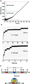
The amount of DNA released from nucleosomes upon disruption. (A) Amount of naked DNA as a function of time derived from data shown in Fig. 1B. The top red dotted line is a comparison with a full-length naked DNA. At higher force, the curves show 17 steps, which correspond to the 17 disruption peaks in Fig. 1B. (B) Step size measurement using a force clamp. The graphs are plots of DNA extension vs. time under constant force. Two examples of the measurements are shown corresponding to two different forces. (C) Map of the critical DNA–histone interactions within an NCP. (Upper) Spatial map of the strongest DNA–histone interaction regions along DNA associated with the histone octamer (16). (Lower) Cartoon representation of a partially disrupted core particle.
Figure 3
Figure 3
Nucleosome reassembly after disruption by repetitive stretching with a velocity clamp. Nucleosomes were repetitively stretched with a 10-s relaxation period after each stretch. (A) Maximum force at ≈50 pN. Force-extension curves of a nucleosomal array repetitively stretched three times with maximum force at ≈50 pN: first stretch (black), second stretch (red), and third stretch (blue). (B) Maximum force at ≈60 pN. A nucleosomal array first was stretched to a maximum force of 50 pN (black). In subsequent stretches (red then blue), the maximum force was increased to ≈60 pN.
Figure 4
Figure 4
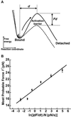
Investigation of the strong interactions at ±40 bp by DFS. (A) Energy diagram illustrating the basic principle of DFS. The effect of an external stretching force exerted along the reaction coordinate is to lower the height of the activation barrier between the bound and detached states. (B) A plot of most probable force F* vs. ln[1/N dF/dt] and its linear fit.
Figure 5
Figure 5
A three-stage model for the mechanical disruption of the NCP. See text for a description.

Comment in

Similar articles

Cited by

References

    1. Kornberg R, Thomas J. Science. 1974;184:865–868. - PubMed
    1. Luger K, Mader A, Richmond R, Sargent D, Richmond T. Nature (London) 1997;389:251–260. - PubMed
    1. Anderson J, Widom J. J Mol Biol. 2000;296:979–987. - PubMed
    1. Furrer P, Bednar J, Dubochet J, Hamiche A, Prunell A. J Struct Biol. 1995;114:177–183. - PubMed
    1. Cui Y, Bustamante C. Proc Natl Acad Sci USA. 2000;97:127–132. - PMC - PubMed

Publication types