. 2002 Feb 19;99(4):1984-9.
doi: 10.1073/pnas.042689299.

Functional studies on the role of the C-terminal domain of mammalian polo-like kinase

Affiliations

Functional studies on the role of the C-terminal domain of mammalian polo-like kinase

Young-Joo Jang et al. Proc Natl Acad Sci U S A. .

Abstract

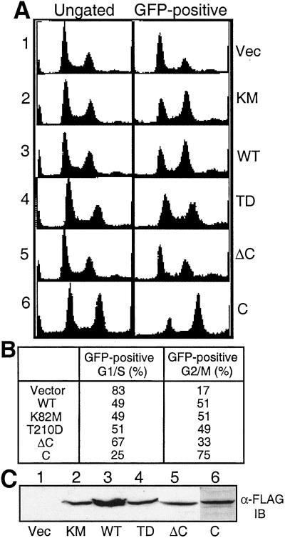
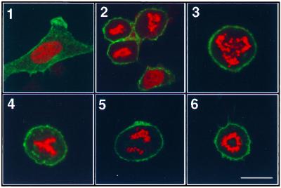
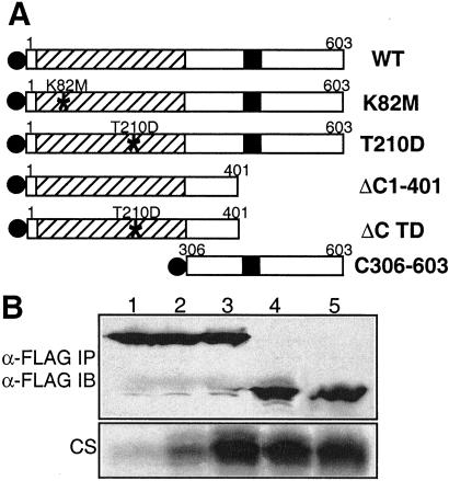
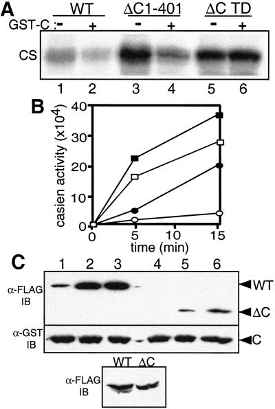
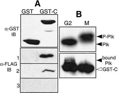
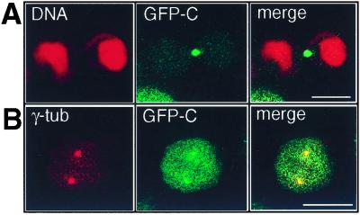
Mammalian polo-like kinase (Plk) acts at various stages in early and late mitosis. Plk is phosphorylated and activated in mitosis, and the proper subcellular localization of Plk is essential for mitotic regulation. We have observed that overexpression of the C-terminal domain of Plk is more effective than wild-type or kinase-defective Plk in causing mitotic delay or arrest. The specific activity of Plk with C-terminal deletions or substitution of aspartate for threonine-210 is increased severalfold relative to wild type. We show in this communication that the C-terminal domain can bind to full-length or the catalytic domain of Plk and inhibit its kinase activity, and that this binding is disrupted when threonine-210 is substituted with an aspartic acid residue. The C-terminal domain binds unphosphorylated Plk from G(2) arrested cells, but not phosphorylated Plk from mitotic cells. Green fluorescent protein-C-terminal Plk is localized at the centrosome and the midbody of transfected cells as shown previously for full-length enzyme. These and other data indicate that although the C terminus serves to regulate Plk kinase activity, the localization of the C terminus at the centrosome and other sites in transfected cells may block the correct localization of endogenous Plk.


Figures

Figure 1
Figure 1
Transient expression of the C-terminal domain in HeLa cells induces mitotic arrest. (A) HeLa cells were cotransfected with pEGFP-F and various pCMV-FLAG-Plk constructs and fixed as described in Materials and Methods. DNA profiles of GFP-positive cells were determined by fluorescence-activated cell sorter (FACS) analysis. Left (ungated) and Right (GFP-positive) represent the DNA profiles from total cell populations and GFP-positive transfected cells after gating, respectively. 1, DNA profiles from transfectants with pCMV-FLAG vector; 2, with pCMV-FLAG-Plk K82M; 3, with pCMV-FLAG-Plk WT; 4, with pCMV-FLAG-Plk T210D; 5, with pCMV-FLAG-ΔC1–401; 6, with pCMV-FLAG-C306–603. (B) The percent of G2/M phase cells determined from the GFP-positive population by ModFit analysis. Data represent three independent experiments. (C) Western blot analysis of expressed proteins from transfected cell lysates with anti-FLAG antibody.
Figure 2
Figure 2
Expression of the C-terminal domain induces accumulation of cells with mitotic phenotypes. (A) A membrane-bound EGFP (green) expression construct was used as the transfection marker, and the DNA was labeled with propidium iodide (red) after transfection as previously described. Most of the cells transfected with control vector are in interphase (1). Cells transfected with the C306–603 construct accumulate in M-phase (2). In some transfected mitotic cells, chromosomes appear to be randomly distributed (3), misaligned (4), or unevenly segregated (5). The circular chromosome is similar to the phenotype first observed in Drosophila polo mutants (6). (Scale bar, 15 μm.)
Figure 3
Figure 3
Schematic representation of Plk1 proteins and kinase activity of immunocomplexes. (A) Plk wild-type and mutant proteins used in this study. The catalytic domain and polo-box are represented by deviant lined box and filled box, respectively. FLAG epitopes on the N-termini are shown as filled circles. From top to bottom: full-length wild-type Plk (WT); kinase-defective mutant Plk (K82M); Plk mutated Thr-210 to Asp (T210D); Plk deleted of the C-terminal amino acids 402–603 (ΔC1–401); N-terminal domain containing the mutation, T210D (ΔC TD); Plk deleted of the N-terminal amino acids 1–305 (C306–603). (B) Kinase activities of Plk proteins immunoprecipitated from transfected HeLa cell lysates. The amounts of immunoprecipitated FLAG-Plk proteins were detected with anti-FLAG antibody (Upper). The kinase activity of FLAG-Plk was measured by incubation with casein for 30 min as described in Materials and Methods (Lower, CS). 1, FLAG-Plk K82M; 2, FLAG-Plk WT; 3, FLAG-Plk T210D; 4, FLAG-N terminus Plk (ΔC1–401); 5, FLAG-N terminus Plk containing T210D mutation (ΔC TD).
Figure 4
Figure 4
The C-terminal domain of Plk inhibits its kinase activity. (A) Inhibitory effect of the C-terminal domain. In in vitro kinase assays, immunoprecipitated FLAG-Plk WT and FLAG-N-terminal domains from transfected HeLa cells were incubated with or without 1–2 μg of C-terminal domain protein C306–603 (GST-C), which was purified from High Five (Hi5) cells. The Plk activity was measured with casein as a substrate (CS). 1, 3, and 5, incubated without GST-C; 2, 4, and 6, incubated with GST-C. (B) Time course of the wild-type and N-terminal Plk kinase activity. Purified wild-type and N-terminal Plk proteins from Hi5 cells were preincubated with or without an equal molar level of purified C-terminal protein for 20 min at 30°C, and then assayed for protein kinase activity. The reactions were stopped by adding SDS/PAGE sample buffer. The samples were boiled and resolved by PAGE. The casein bands were excised from the gel and the radioactivity was quantitated by liquid scintillation spectrometry. Filled squares, ΔC1–401 protein; open squares, ΔC1–401 with C306–603; filled circles, Plk WT; open circles, Plk WT with C306–603. (C) Binding of the C terminus to wild-type and N-terminal Plk. GST-fused C-terminal construct, expressed in Hi5 cells, was incubated with HeLa cell lysates, which had been transfected with FLAG-tagged Plk constructs. After incubation for various times, GST-fused proteins were precipitated with glutathione-agarose beads. 1 and 4, incubation for 5 min; 2 and 5, incubation for 30 min; 3 and 6, incubation for 2 h. The wild-type (–3) and N-terminal (–6) Plk proteins associated with the GST-C-terminal domain were detected by Western blot analysis (α-FLAG IB). After stripping the FLAG signal, the membrane was incubated with GST antibody to quantify the GST-C protein (α-GST IB). The levels of wild-type and N-terminal Plk (WT and ΔC) in 10% of total lysates were analyzed by Western analysis (α-FLAG IB).
Figure 5
Figure 5
The C-terminal domain of Plk binds to Plk, and does not bind to phosphorylated mitotic Plk. (A) GST-fused C-terminal protein C306–603 (GST-C) purified from High Five cells was incubated with lysates of HeLa cells that had been transfected with various FLAG-tagged Plk constructs. 1, FLAG-Plk WT protein bound to GST-C; 2, FLAG-ΔC1–401 protein bound to GST-C; 3, FLAG-ΔC1–401 protein (T210D) bound to GST-C. The Plk proteins associated with the GST-C-terminal domain were detected by Western blot analysis with anti-FLAG antibody (α-FLAG IB). The amount of GST or GST-C protein used was detected with anti-GST antibody (α-GST IB). (B) Binding of endogenous Plk to C-terminal domain (GST-C). GST-fused C306–603 protein was incubated with lysates of FT210 cells that were arrested in G2 or mitosis as previously described, and was collected on glutathione-agarose. Endogenous Plks displayed phosphorylated M-phase and dephosphorylated G2 forms (Upper) and endogenous Plk (filled arrowhead) associated with the C306–603 protein (GST-C, opened arrowhead) were detected by Western blot with anti-Plk antibody (Lower). The cell lysates were analyzed on 8% SDS/PAGE gel (acryl:bis = 120:1) (15).
Figure 6
Figure 6
Localization of GFP-Plk C terminus in HeLa cells. HeLa cells were transfected with GFP-C, fixed, and analyzed as described in Materials and Methods. (A) The central midbody is intensely labeled with GFP in a telophase cell (GFP-C, green). The chromosomal DNA is stained with propidium iodide (DNA, red). (B) GFP-C is localized predominantly at centrosomes in a prometaphase cell (GFP-C). γ-tubulin on centrosomes is stained with anti-γ-tubulin antibody. (Scale bars, 15 μm.)

Similar articles

Cited by

References

    1. Morgan D O. Nature (London) 1995;374:131–134. - PubMed
    1. Nigg EA. BioEssays. 1995;17:471–480. - PubMed
    1. Glover D M, Hagan I M, Tavares A M. Genes Dev. 1998;12:3777–3787. - PubMed
    1. Clay F J, McEwen S J, Bertoncello I, Wilks A F, Dunn A R. Proc Natl Acad Sci USA. 1993;90:4882–4886. - PMC - PubMed
    1. Sunkel C E, Glover D M. J Cell Sci. 1988;89:25–38. - PubMed

Publication types

MeSH terms

LinkOut - more resources