

Integration of foreign DNA during natural transformation of Acinetobacter sp. by homology-facilitated illegitimate recombination
- PMID: 11854504
- PMCID: PMC122324
- DOI: 10.1073/pnas.042263399
Integration of foreign DNA during natural transformation of Acinetobacter sp. by homology-facilitated illegitimate recombination
Abstract
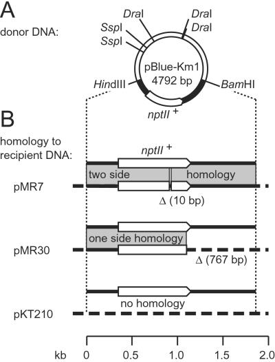
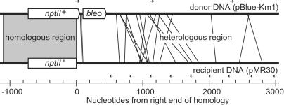
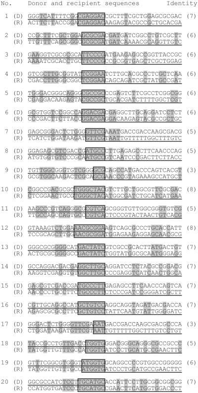
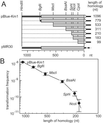
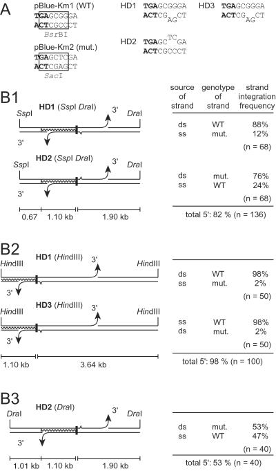
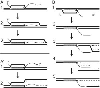
The active uptake of extracellular DNA and its genomic integration is termed natural transformation and constitutes a major horizontal gene-transfer mechanism in prokaryotes. Chromosomal DNA transferred within a species can be integrated effectively by homologous recombination, whereas foreign DNA with low or no sequence homology would rely on illegitimate recombination events, which are rare. By using the nptII(+) gene (kanamycin resistance) as selectable marker, we found that the integration of foreign DNA into the genome of the Gram-negative Acinetobacter sp. BD413 during transformation indeed was at least 10(9)-fold lower than that of homologous DNA. However, integration of foreign DNA increased at least 10(5)-fold when it was linked on one side to a piece of DNA homologous to the recipient genome. Analysis of foreign DNA integration sites revealed short stretches of sequence identity (3-8 bp) between donor and recipient DNA, indicating illegitimate recombination events. These findings suggest that homologous DNA served as a recombinational anchor facilitating illegitimate recombination acting on the same molecule. Homologous stretches down to 183 nucleotides served as anchors. Transformation with heteroduplex DNA having different nucleotide sequence tags in the strands indicated that strands entered the cytoplasm 3' to 5' and that strands with either polarity were integrated by homologous recombination. The process led to the genomic integration of thousands of foreign nucleotides and often was accompanied by deletion of a roughly corresponding length of recipient DNA. Homology-facilitated illegitimate recombination would explain the introgression of DNA in prokaryotic genomes without the help of mobile genetic elements.
Figures
Similar articles
-
The RecJ DNase strongly suppresses genomic integration of short but not long foreign DNA fragments by homology-facilitated illegitimate recombination during transformation of Acinetobacter baylyi.Mol Microbiol. 2007 May;64(3):691-702. doi: 10.1111/j.1365-2958.2007.05692.x. Mol Microbiol. 2007. PMID: 17462017
-
Double illegitimate recombination events integrate DNA segments through two different mechanisms during natural transformation of Acinetobacter baylyi.Mol Microbiol. 2008 Mar;67(5):984-95. doi: 10.1111/j.1365-2958.2007.06096.x. Epub 2008 Jan 8. Mol Microbiol. 2008. PMID: 18194157
-
Mechanisms of homology-facilitated illegitimate recombination for foreign DNA acquisition in transformable Pseudomonas stutzeri.Mol Microbiol. 2003 May;48(4):1107-18. doi: 10.1046/j.1365-2958.2003.03498.x. Mol Microbiol. 2003. PMID: 12753199
-
Molecular aspects of gene transfer and foreign DNA acquisition in prokaryotes with regard to safety issues.Appl Microbiol Biotechnol. 2010 Apr;86(4):1027-41. doi: 10.1007/s00253-010-2489-3. Epub 2010 Feb 27. Appl Microbiol Biotechnol. 2010. PMID: 20191269 Review.
-
Illegitimate DNA integration in mammalian cells.Gene Ther. 2003 Oct;10(21):1791-9. doi: 10.1038/sj.gt.3302074. Gene Ther. 2003. PMID: 12960968 Review.
Cited by
-
Genetic structures at the origin of acquisition and expression of the carbapenem-hydrolyzing oxacillinase gene blaOXA-58 in Acinetobacter baumannii.Antimicrob Agents Chemother. 2006 Apr;50(4):1442-8. doi: 10.1128/AAC.50.4.1442-1448.2006. Antimicrob Agents Chemother. 2006. PMID: 16569863 Free PMC article.
-
Spread of novel aminoglycoside resistance gene aac(6')-Iad among Acinetobacter clinical isolates in Japan.Antimicrob Agents Chemother. 2004 Jun;48(6):2075-80. doi: 10.1128/AAC.48.6.2075-2080.2004. Antimicrob Agents Chemother. 2004. PMID: 15155202 Free PMC article.
-
Strand-biased gene distribution in bacteria is related to both horizontal gene transfer and strand-biased nucleotide composition.Genomics Proteomics Bioinformatics. 2012 Aug;10(4):186-96. doi: 10.1016/j.gpb.2012.08.001. Epub 2012 Aug 8. Genomics Proteomics Bioinformatics. 2012. PMID: 23084774 Free PMC article.
-
Biofilms preserve the transmissibility of a multi-drug resistance plasmid.NPJ Biofilms Microbiomes. 2022 Dec 9;8(1):95. doi: 10.1038/s41522-022-00357-1. NPJ Biofilms Microbiomes. 2022. PMID: 36481746 Free PMC article.
-
Homology-dependent DNA transfer from plants to a soil bacterium under laboratory conditions: implications in evolution and horizontal gene transfer.Transgenic Res. 2003 Aug;12(4):425-37. doi: 10.1023/a:1024387510243. Transgenic Res. 2003. PMID: 12885164
References
Publication types
MeSH terms
Substances
LinkOut - more resources
Full Text Sources
Other Literature Sources
