. 2002 Feb 19;99(4):2199-204.
doi: 10.1073/pnas.042678299.

Marrow stromal cells form guiding strands in the injured spinal cord and promote recovery

Affiliations

Marrow stromal cells form guiding strands in the injured spinal cord and promote recovery

C P Hofstetter et al. Proc Natl Acad Sci U S A. .

Abstract

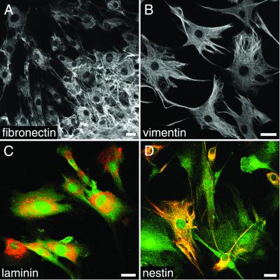
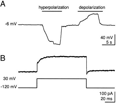
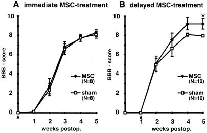
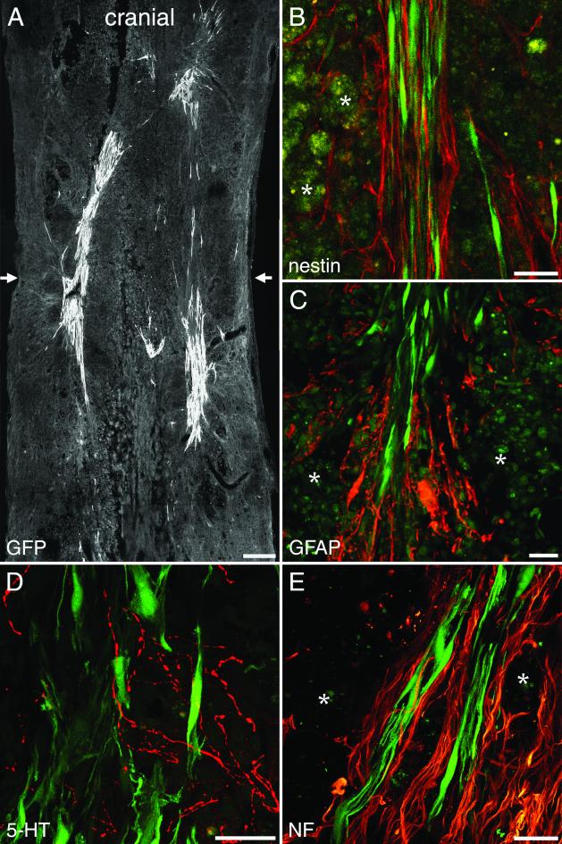
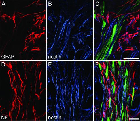
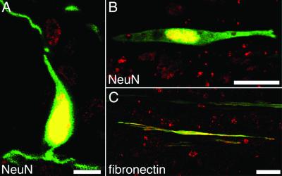
Marrow stromal cells (MSC) can be expanded rapidly in vitro and differentiated into multiple mesodermal cell types. In addition, differentiation into neuron-like cells expressing markers typical for mature neurons has been reported. To analyze whether such cells, exposed to differentiation media, could develop electrophysiological properties characteristic of neurons, we performed whole-cell recordings. Neuron-like MSC, however, lacked voltage-gated ion channels necessary for generation of action potentials. We then delivered MSC into the injured spinal cord to study the fate of transplanted MSC and possible effects on functional outcome in animals rendered paraplegic. MSC given 1 week after injury led to significantly larger numbers of surviving cells than immediate treatment and significant improvements of gait. Histology 5 weeks after spinal cord injury revealed that MSC were tightly associated with longitudinally arranged immature astrocytes and formed bundles bridging the epicenter of the injury. Robust bundles of neurofilament-positive fibers and some 5-hydroxytryptamine-positive fibers were found mainly at the interface between graft and scar tissue. MSC constitute an easily accessible, easily expandable source of cells that may prove useful in the establishment of spinal cord repair protocols.


Figures

Figure 1
Figure 1
Appearance of MSC in culture. (A) All MSC display fibronectin-IR during culture conditions. Extensive deposition of fibronectin is observed in the cell cluster in the lower right corner. Vimentin-IR (B) and laminin-IR (C) are expressed by all MSC. (D) Nestin-IR is detected in a subset of MSC with different morphologies. In C and D, the GFP cell marker (green) is shown together with laminin or nestin (red). (Bars = 25 μm.)
Figure 2
Figure 2
Electrophysiological properties of a neuron-like MSC. (A) Membrane potential of a neuron-like MSC at rest and during hyperpolarization and depolarization. Note that no action potentials were elicitable even after hyperpolarization to reactivate possible voltage-gated ion channels. (B) Voltage-gated currents elicited via a voltage command stepping from −120 mV to 30 mV. Neither voltage-gated Na+ channels nor voltage-gated K+ channels are present.
Figure 3
Figure 3
(A) Analysis of locomotor recovery as measured by BBB scores. Animals treated with MSC immediately after SCI do not differ from control animals. (B) Delayed MSC treatment significantly improved locomotor recovery. *, P = 0.013. Data represent means ± SEM. Arrowheads indicate the treatment time.
Figure 4
Figure 4
One-week-delayed transplantation of MSC after SCI. (A) MSC form bundles bridging the epicenter of the lesion visualized by the transgenic GFP marker. Arrows indicate the location of the impact injury. (B) Nestin-immunoreactive immature astrocytes with longitudinally aligned processes (red) are found within MSC bundles (green). (C) GFAP-IR (red) marks astrocytic processes penetrating the grafted cell aggregates (green). (D) 5-HT-positive fibers (red) are present among the implanted MSC (green). (E) Robust NF-IR nerve fiber bundles (red) are found at the interface between MSC and host tissue. (B, C, and E) Asterisks indicate macrophages. [Bars = 250 μm (A) and 25 μm (BE).]
Figure 5
Figure 5
MSC bundles guide host neuropil. (AC) Beyond the astrocytic scar surrounding the epicenter of the lesion (A), nestin-positive (blue) and GFAP-negative immature astrocytes (B) are found closely associated with transplanted MSC (C). (DF) Neurofilament-IR fibers (red) are found in close relationship with nestin-IR fibers (blue) mainly in the periphery of MSC bundles. (Bars = 25 μm.)
Figure 6
Figure 6
(A and B) Five weeks after SCI, MSC express a distinct nuclear NeuN-IR. (C) All MSC are fibronectin-positive. [Bar = 10 μm (A) and 25 μm (B and C).]

References

    1. Fawcett J W, Asher R A. Brain Res Bull. 1999;49:377–391. - PubMed
    1. Skene J H. Annu Rev Neurosci. 1989;12:127–156. - PubMed
    1. Widenfalk J, Lundstromer K, Jubran M, Brene S, Olson L. J Neurosci. 2001;21:3457–3475. - PMC - PubMed
    1. Schwartz M. Prog Brain Res. 2000;128:259–263. - PubMed
    1. Schnell L, Schwab M E. Nature (London) 1990;343:269–272. - PubMed

Publication types

MeSH terms

Substances