

Molecular specialization of breast vasculature: a breast-homing phage-displayed peptide binds to aminopeptidase P in breast vasculature
- PMID: 11854520
- PMCID: PMC122351
- DOI: 10.1073/pnas.251687998
Molecular specialization of breast vasculature: a breast-homing phage-displayed peptide binds to aminopeptidase P in breast vasculature
Abstract
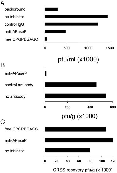
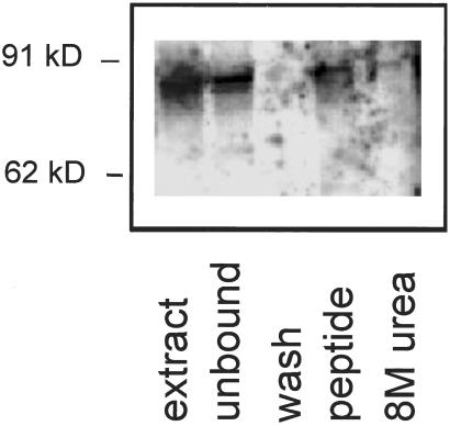
In vivo phage display identifies peptides that selectively home to the vasculature of individual organs, tissues, and tumors. Here we report the identification of a cyclic nonapeptide, CPGPEGAGC, which homes to normal breast tissue with a 100-fold selectivity over nontargeted phage. The homing of the phage is inhibited by its cognate synthetic peptide. Phage localization in tissue sections showed that the breast-homing phage binds to the blood vessels in the breast, but not in other tissues. The phage also bound to the vasculature of hyperplastic and malignant lesions in transgenic breast cancer mice. Expression cloning with a phage-displayed cDNA library yielded a phage that specifically bound to the breast-homing peptide. The cDNA insert was homologous to a fragment of aminopeptidase P. The homing peptide bound aminopeptidase P from malignant breast tissue in affinity chromatography. Antibodies against aminopeptidase P inhibited the in vitro binding of the phage-displayed cDNA to the peptide and the in vivo homing of phage carrying the peptide. These results indicate that aminopeptidase P is the receptor for the breast-homing peptide. This peptide may be useful in designing drugs for the prevention and treatment of breast cancer.
Figures
Similar articles
-
Identification of a peptide sequence targeting mammary vasculature via RPLP0 during lactation.Peptides. 2010 Dec;31(12):2247-54. doi: 10.1016/j.peptides.2010.09.008. Epub 2010 Sep 21. Peptides. 2010. PMID: 20863866
-
Targeting of tumor blood vessels: a phage-displayed tumor-homing peptide specifically binds to matrix metalloproteinase-2-processed collagen IV and blocks angiogenesis in vivo.Mol Cancer Res. 2009 Jul;7(7):1078-85. doi: 10.1158/1541-7786.MCR-08-0538. Epub 2009 Jul 7. Mol Cancer Res. 2009. PMID: 19584266
-
Identification of synovium-specific homing peptides by in vivo phage display selection.Arthritis Rheum. 2002 Aug;46(8):2109-20. doi: 10.1002/art.10464. Arthritis Rheum. 2002. PMID: 12209516
-
Vascular zip codes in angiogenesis and metastasis.Biochem Soc Trans. 2004 Jun;32(Pt3):397-402. doi: 10.1042/BST0320397. Biochem Soc Trans. 2004. PMID: 15157146 Review.
-
Identification and Characterization of Homing Peptides Using In Vivo Peptide Phage Display.Methods Mol Biol. 2015;1324:205-22. doi: 10.1007/978-1-4939-2806-4_14. Methods Mol Biol. 2015. PMID: 26202272 Review.
Cited by
-
Anti-angiogenic peptides application in cancer therapy; a review.Res Pharm Sci. 2021 Oct 15;16(6):559-574. doi: 10.4103/1735-5362.327503. eCollection 2021 Dec. Res Pharm Sci. 2021. PMID: 34760005 Free PMC article. Review.
-
Navigare necessere est. Improved navigation would help to solve two crucial problems in modern drug therapy: toxicity and precise delivery.EMBO Rep. 2005 Aug;6(8):695-700. doi: 10.1038/sj.embor.7400484. EMBO Rep. 2005. PMID: 16065058 Free PMC article.
-
Improved Antiglioblastoma Activity and BBB Permeability by Conjugation of Paclitaxel to a Cell-Penetrative MMP-2-Cleavable Peptide.Adv Sci (Weinh). 2020 Dec 21;8(3):2001960. doi: 10.1002/advs.202001960. eCollection 2021 Feb. Adv Sci (Weinh). 2020. PMID: 33552853 Free PMC article.
-
In vivo selection of tumor-targeting RNA motifs.Nat Chem Biol. 2010 Jan;6(1):22-4. doi: 10.1038/nchembio.277. Epub 2009 Nov 29. Nat Chem Biol. 2010. PMID: 19946274 Free PMC article.
-
Tumor-targeting peptides from combinatorial libraries.Adv Drug Deliv Rev. 2017 Feb;110-111:13-37. doi: 10.1016/j.addr.2016.05.009. Epub 2016 May 19. Adv Drug Deliv Rev. 2017. PMID: 27210583 Free PMC article. Review.
References
-
- Ruoslahti E, Rajotte D. Annu Rev Immunol. 2000;18:813–827. - PubMed
-
- Streeter P R, Berg E L, Rouse B T N, Bargatze R F, Butcher E C. Nature (London) 1988;331:41–46. - PubMed
-
- Johnson R C, Augustin-Voss H G, Zhu D, Pauli B U. Cancer Res. 1991;51:394–399. - PubMed
-
- Pasqualini R, Ruoslahti E. Nature (London) 1996;380:360–364. - PubMed
Publication types
MeSH terms
Substances
Grants and funding
LinkOut - more resources
Full Text Sources
Other Literature Sources
