Case Reports
. 2002 Apr;70(4):1015-22.
doi: 10.1086/339518. Epub 2002 Feb 20.

Premature chromosome condensation in humans associated with microcephaly and mental retardation: a novel autosomal recessive condition

Affiliations
Case Reports

Premature chromosome condensation in humans associated with microcephaly and mental retardation: a novel autosomal recessive condition

Heidemarie Neitzel et al. Am J Hum Genet. 2002 Apr.

Abstract

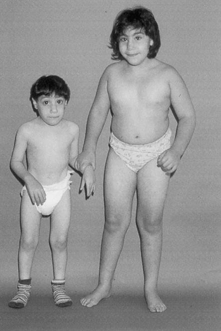
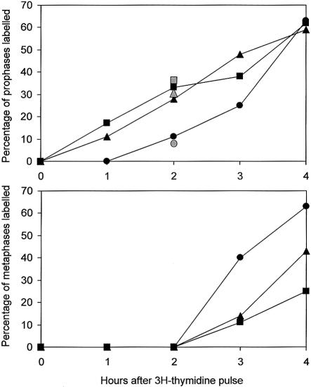
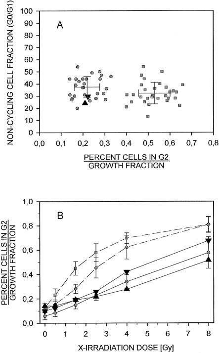
We report a novel autosomal recessive disorder characterized by premature chromosome condensation in the early G2 phase. It was observed in two siblings, from consanguineous parents, affected with microcephaly, growth retardation, and severe mental retardation. Chromosome analysis showed a high frequency of prophase-like cells (>10%) in lymphocytes, fibroblasts, and lymphoblast cell lines with an otherwise normal karyotype. (3)H-thymidine-pulse labeling and autoradiography showed that, 2 h after the pulse, 28%-35% of the prophases were labeled, compared with 9%-11% in healthy control subjects, indicating that the phenomenon is due to premature chromosome condensation. Flow cytometry studies demonstrate that the entire cell cycle is not prolonged, compared with that in healthy control subjects, and compartment sizes did not differ from those in healthy control subjects. No increased reaction of the cells to X-irradiation or treatments with the clastogens bleomycin and mitomycin C was observed, in contrast to results in the cell-cycle mutants ataxia telangiectasia and Fanconi anemia. The rates of sister chromatid exchanges and the mitotic nondisjunction rates were inconspicuous. Premature entry of cells into mitosis suggests that a gene involved in cell-cycle regulation is mutated in these siblings.


Figures

Figure  1
Figure 1
The older and the younger sibling at the ages of 7 and 5 years, respectively.
Figure  2
Figure 2
Premature chromosome condensation in lymphocytes and fibroblasts. a, Chromosome preparation from PHA-stimulated 72-h lymphocyte culture harvested without prior colcemid treatment. b, Chromosomes in prophase nuclei of a fibroblast line, showing an intact nuclear membrane, as observed in most of the prophase-like cells. c and d, Normal alignment of the chromosomes in metaphase plates from fibroblasts. e, Normal anaphase lagging in the fibroblasts harvested without prior colcemid treatment. f, Normal segregation of homologous chromosomes in lymphoblast cells analyzed after blocking of the cytokinesis with cytochalasin and subsequent FISH performed with two centromeric probes from chromosome 8 and chromosome 18. g, Two cells after Hoechst staining of the prematurely condensed chromosomes (arrows). h, The same cells after indirect immunostaining with lamin B antibodies, demonstrating the intact nuclear membrane.
Figure  3
Figure 3
Cell-cycle analysis after 3H-thymidine-pulse-labeling of logarithmically growing lymphoblast cell lines from both siblings. The cells were treated for 10 min with 1 μCi/ml 3H-thymidine (104, 7 Ci/mmol; specific activity: 104 Ci/mM) and were harvested for chromosome preparation at 1-h intervals. The numbers of labeled and unlabeled pro- and metaphase cells was determined after autoradiography for patient 1 (triangle), patient 2 (square), and a control (circle). Gray symbols give the percentage determined in an independent second experiment 2 h after pulse labeling.
Figure  4
Figure 4
Normal response of the lymphoblast cell lines from patient 1 (upright triangle) and patient 2 (inverted triangle) to ionizing radiation. A, The G2-phase proportions of these lines, relative to their corresponding growth fractions at 1.5 Gy of irradiation, were 0.21 and 0.22, respectively. These ratios were within the range of 28 normal control lines (mean ± 1 SD = 0.22 ± 0.06; range 0.14–0.33), as opposed to 30 radiosensitive A-T lines (mean ± 1 SD = 0.53 ± 0.07; range 0.40–0.66). All cultures revealed similar growth fractions (noncycling cells: patient 1, 24%; patient 2, 30%; mean ± 1 SD of the healthy control subjects: 37.2% ± 8.7%, range: 20%–54%; of the A-T lines: 32.7 ± 8.9 %, range: 13%–54%). B, The G2 phase proportions, relative to the corresponding growth fractions of the lines of both patients at a broader range of irradiation (0.5–8.0 Gy) resemble the dose-response curve of eight healthy control subjects (blackened circles, mean ± 1 SD) but are distinct from those of five A-T lines (blackened squares, mean ± 1 SD) and of three NBS lines (unblackened circles, mean ± 1 SD).
Figure  5
Figure 5
Normal response of the cells from patient 1 (upright triangle) and patient 2 (inverted triangle) to MMC. A, The G2-phase proportion of primary lymphocytes of patient 2, relative to their corresponding growth fraction at 10 ng/ml MMC, was 0.30. This ratio was within the limits of 23 normal control lymphocyte cultures (mean ± 1 SD = 0.25 ± 0.05; range 0.18–0.34), as opposed to 35 MMC-sensitive FA cultures (mean ± 1 SD = 0.63 ± 0.06; range 0.47–0.73). All cultures revealed similar growth fractions (percent noncycling cells, patient 2: 40%; mean ± 1 SD of the healthy control subjects 31.8 ± 13.1%; range 13.4%–71.2%; mean ± 1 SD of the FA lymphocytes: 39.7% ± 15.7%; range 13.4%–81.1%). B, The G2-phase proportions of the lymphoblast lines of both patients relative to their corresponding growth fractions at 10 ng/ml MMC were 0.24 and 0.27, respectively. These ratios were within the range of 12 normal control lines (mean ± 1 SD = 0.23 ± 0.04; range 0.18–0.31) as opposed to 5 MMC-sensitive FA lines (mean ± 1 SD = 0.45 ± 0.04; range 0.41–0.51). The lines of both patients revealed similar growth fractions (percent noncycling cells: patient 1, 15.9%; patient 2, 18.1%) as the normal control lines (mean ± 1 SD = 23.0% ± 8.9%; range 4.7%–38.5%).

Similar articles

Cited by

References

Electronic-Database Information

    1. Online Mendelian Inheritance in Man (OMIM), http://www.ncbi.nlm.nih.gov/Omim/ (for RCC1 [MIM 179710], CDC25A [MIM 116947], CDC25B [MIM 116949], CDC25C [MIM 157680], cyclin B1 [MIM 123836], cyclin A2 [MIM 123835], CDK1 [MIM 116940], and CDK2 [MIM 116953])

References

    1. Furuno N, den Elzen N, Pines J (1999) Human cyclin A is required for mitosis until mid prophase. J Cell Biol 147:295–306 - PMC - PubMed
    1. Georgatos SD, Pyrpasopoulou A, Theodoropoulos PA (1997) Nuclear envelope breakdown in mammalian cells involves stepwise lamina disassembly and microtubule-drive deformation of the nuclear membrane. J Cell Sci 110:2129–2140 - PubMed
    1. Goldstone S, Pavey S, Forrest A, Sinnamon J, Gabrielli B (2001) Cdc25-dependent activation of cyclin A/cdk2 is blocked in G2 phase arrested cells independently of ATM/ATR. Oncogene 20:921–932 - PubMed
    1. Hagting A, Jackman M, Simpson K, Pines J (1999) Translocation of cyclin B1 to the nucleus at prophase requires a phosphorylation-dependent nuclear import signal. Curr Biol 9:680–689 - PubMed
    1. Johnson RT, Rao PN (1970) Mammalian cell fusion: induction of premature chromosome condensation in interphase nuclei. Nature 226:717–722 - PubMed

Publication types

MeSH terms

Associated data