. 2001 Dec;17(3):285-92.
doi: 10.1385/JMN:17:3:285.

Roles of BCL-2 and caspase 3 in the adenosine A3 receptor-induced apoptosis

Affiliations

Roles of BCL-2 and caspase 3 in the adenosine A3 receptor-induced apoptosis

E Appel et al. J Mol Neurosci. 2001 Dec.

Abstract

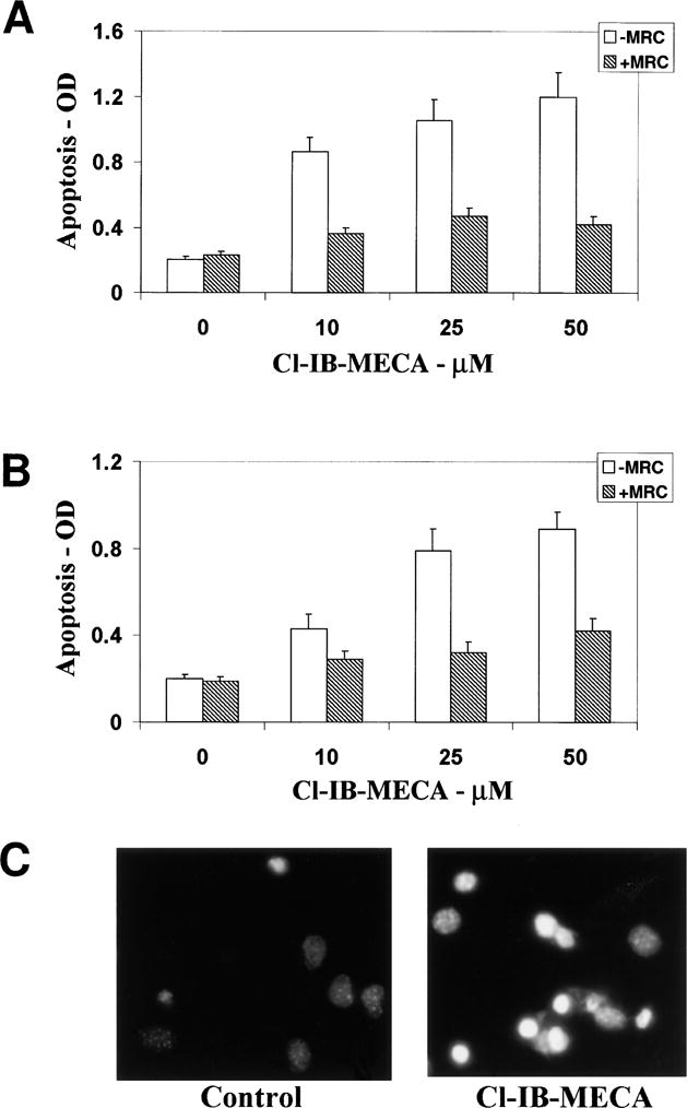
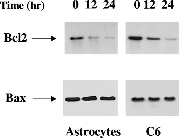
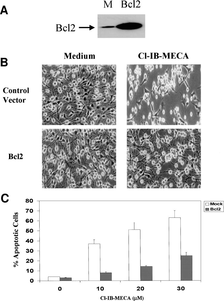
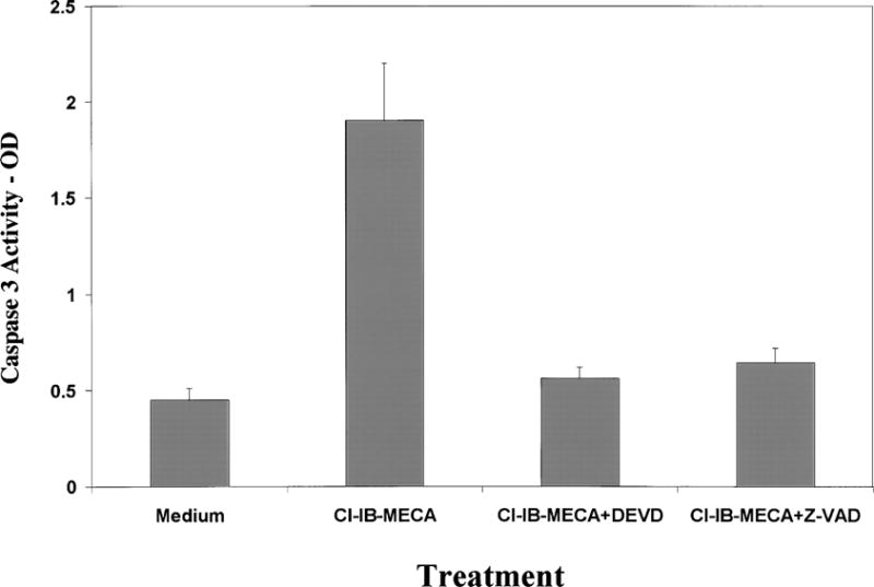
Selective A3 adenosine receptor agonists have been shown to induce apoptosis in a variety of cell types. In this study we examined the effects of adenosine receptor agonists selective for A1, A2A, or A3 receptors on the induction of apoptosis in primary cultures of rat astrocytes and in C6 glial cells. Treatment of the cells with the A3 receptor agonist Cl-IB-MECA (10 microM) induced apoptosis in both cell types. The effects of Cl-IB-MECA were partially antagonized by the A3 receptor-selective antagonist MRS 1191. In contrast, the A1 and A2A receptor agonists, CPA and CGS 21680, respectively, did not have significant effects on apoptosis in these cells. Cl-IB-MECA reduced the expression of endogenous Bcl-2, whereas it did not affect the expression of Bax. Overexpression of Bcl-2 in C6 cells abrogated the induction of apoptosis induced by the A3 agonist. Cl-IB-MECA also induced an increase in caspase 3 activity and caspase inhibitors decreased the apoptosis induced by the A3 agonist. These findings suggest that intense activation of the A3 receptor is pro-apoptotic in glial cells via bcl2 and caspase-3 dependent pathways.


Figures

Fig. 1
Fig. 1
Effects of the A3 receptor agonist Cl-IB-MECA on apoptosis in C6 glial cells. C6 cells (A) and astrocytes (B) were treated with different concentrations of Cl-IB-MECA for 48 h in the presence and absence of the A3 antagonist MRS 1191 (10 μM). Apoptosis was measured by ELISA using anti-histone antibodies. Fragmented DNA was extracted from the cells and was incubated in 96-well plates coated with anti-histone antibodies for 2 h followed by incubation with anti-DNA antibodies conjugated to peroxidase for an additional 2 h. Absorbance was measured at 405 nm. Apoptosis of primary astrocytes was also measured using Hoechst staining (C). Astrocytes were treated with 50 μM Cl-IB-MECA for 48 h and were stained with Hoechst as described in Methods. Results represent the means ± SD of three separate experiments.
Fig. 2
Fig. 2
Effect of the A3 receptor agonist Cl-IB-MECA on the expression of Bcl-2 and Bax. Cells were treated with Cl-IB-MECA (25 μM) for 12 and 24 h. The cells were then harvested and immunoprecipitation of Bcl-2 was performed as described in the Methods or cells were extracted for Western-blot analysis. Following SDS-PAGE, membranes were stained with anti-Bcl-2 antibody or with anti-Bax antibody. The results represent one of three separate experiments, which gave similar results.
Fig. 3
Fig. 3
Effect of Bcl-2 overexpression on the apoptosis induced by the A3 receptor agonist Cl-IB-MECA. Stable transfectants of Bcl-2 or control vector were harvested and subjected to SDS-PAGE and Western blot analysis. Membranes were probed with anti-Bcl-2 antibody (A). Cells overexpressing Bcl2 or empty vector were treated with Cl-IB-MECA for 48 h and the morphology of the cells was examined using a phase-contrast light microscope (B). Cell apoptosis was determined using Hoechst staining and the apoptotic cells were viewed and counted under UV illumination (C). The results represent means ± SD from three separate experiments.
Fig. 4
Fig. 4
Caspase 3 activity. Primary astrocytes or C6 cells treated with 25 μM for 12 h and the activity of caspase 3 (CPP32) was measured using spectrophotometric detection of the cleaved product of the labeled substrate DEVD-p-nitroanilide. The results represent the means ± SD from three separate experiments.

Similar articles

Cited by

References

    1. Abbracchio MP, Rainaldi G, Giammarioli AM, Ceruti S, Brambilla R, Cattabeni F, et al. The A3 adenosine receptor mediates cell spreading, reorganization of actin cytoskeleton, and distribution of Bcl-XL: studies in human astroglioma cells. Biochem Biophys Res Commun. 1997;241:297–304. - PMC - PubMed
    1. Brodie C, Blumberg PM, Jacobson KA. Activation of the A2A adenosine receptor inhibits nitric oxide production in glial cells. FEBS Lett. 1998a;429:139–142. - PMC - PubMed
    1. Brodie C, Bogi K, Acs P, Lorenzo PS, Baskin L, Blumberg PM. Protein kinase C delta (PKCdelta) inhibits the expression of glutamine synthetase in glial cells via the PKCdelta regulatory domain and its tyrosine phosphorylation. J Biol Chem. 1998b;273:30,713–30,718. - PubMed
    1. Brodie C, Weizman N, Katzoff A, Lustig S, Kobiler D. Astrocyte activation by Sindbis virus: expression of GFAP, cytokines, and adhesion molecules. Glia. 1997;19:275–285. - PubMed
    1. Ceruti S, Barbieri D, Franceschi C, Giammarioli AM, Rainaldi G, Malorni W, et al. Effects of adenosine A3 receptor agonists on astrocytes: induction of cell protection at low and cell death at high concentration. Drug Dev Res. 1996;37:177.

MeSH terms