. 2002:3:4.
doi: 10.1186/1471-2121-3-4. Epub 2002 Feb 8.

Quantitation of the distribution and flux of myosin-II during cytokinesis

Affiliations

Quantitation of the distribution and flux of myosin-II during cytokinesis

Douglas N Robinson et al. BMC Cell Biol. 2002.

Abstract

Background: During cytokinesis, the cell's equator contracts against the cell's global stiffness. Identifying the biochemical basis for these mechanical parameters is essential for understanding how cells divide. To achieve this goal, the distribution and flux of the cell division machinery must be quantified. Here we report the first quantitative analysis of the distribution and flux of myosin-II, an essential element of the contractile ring.

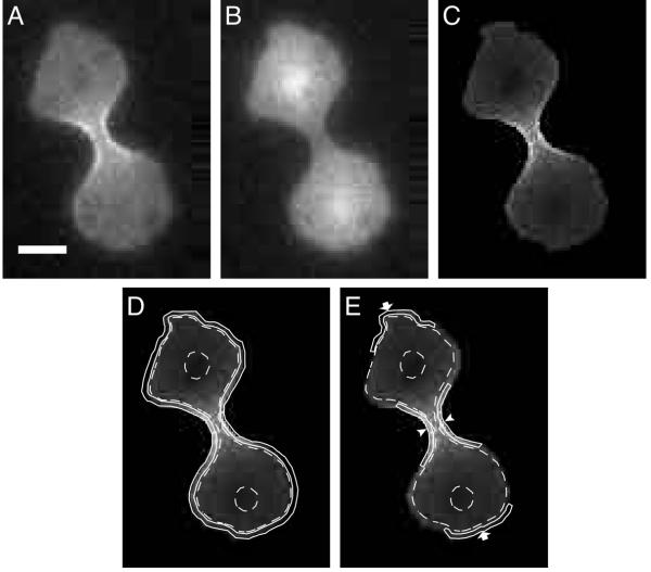
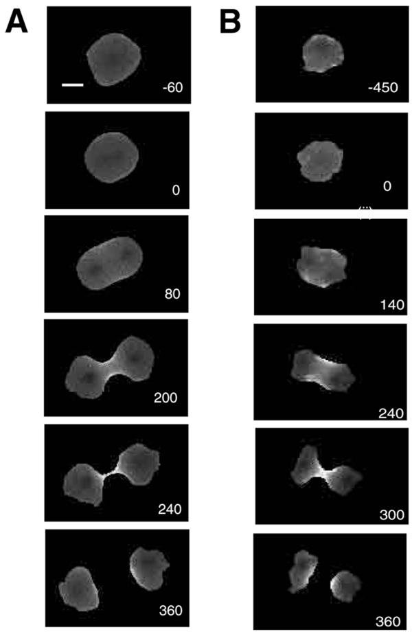
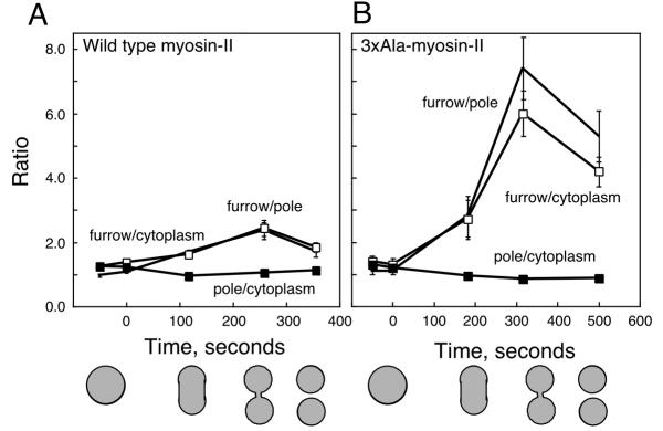
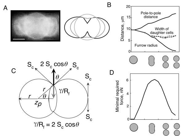
Results: The fluxes of myosin-II in the furrow cortex, the polar cortex, and the cytoplasm were examined using ratio imaging of GFP fusion proteins expressed in Dictyostelium. The peak concentration of GFP-myosin-II in the furrow cortex is 1.8-fold higher than in the polar cortex and 2.0-fold higher than in the cytoplasm. The myosin-II in the furrow cortex, however, represents only 10% of the total cellular myosin-II. An estimate of the minimal amount of this motor needed to produce the required force for cell cleavage fits well with this 10% value. The cell may, therefore, regulate the amount of myosin-II sent to the furrow cortex in accordance with the amount needed there. Quantitation of the distribution and flux of a mutant myosin-II that is defective in phosphorylation-dependent thick filament disassembly confirms that heavy chain phosphorylation regulates normal recruitment to the furrow cortex.

Conclusion: The analysis indicates that myosin-II flux through the cleavage furrow cortex is regulated by thick filament phosphorylation. Further, the amount of myosin-II observed in the furrow cortex is in close agreement with the amount predicted to be required from a simple theoretical analysis.


Figures

Figure 1
Figure 1
Regions defined for analysis. A. A dividing cell is imaged with excitation at 490 nm, showing GFP-myosin-II distribution. B. The same cell is imaged with excitation at 405 nm, showing the volume marker, NLS-GFP (nuclear localization signal fused to GFP) distribution. C. Division of the image in A with the image in B leads to this resulting ratio image. D. The cortex is defined as the 0.35-μm thick shell of the cell, bordered by the solid continuous lines. The cytoplasm (outlined by dashed lines) is the total volume of the cell minus the volume of the cortex. It should be noted that the fluorescence intensity of the cytoplasm in the central region of the cell included the overlying and underlying cortex. However, the contribution of these cortical components to the total fluorescence intensity of the cytoplasmic region was calculated to be less than 2%. The nuclei are encircled to mark their positions, but their combined volume is inconsequential. E. The furrow cortex region is indicated with arrowheads and the polar cortex is indicated with arrows. Bar, 5 μm.
Figure 2
Figure 2
A. A time series of ratio images of a wild type GFP-myosin-II expressing cell going through cytokinesis. The numbers indicate seconds with 0 defined as the point of nuclear division. B. A time series of ratio images of a 3 × Ala GFP-myosin-II expressing cell going through cytokinesis. Bar, 5 μm.
Figure 3
Figure 3
The relative intensity ratios for the pole cortex, furrow cortex and cytoplasm were measured as a function of time. Zero seconds indicates the point of nuclear division. A. The graph shows the ratios obtained with wild-type GFP-myosin-II. The shapes of the cells at four different time points are indicated by the cartoons. B. This graph shows the ratios obtained with cells expressing 3 × Ala GFP-myosin-II. Standard errors for each time point are based on n≥ 4. Markers: furrow-to-pole, no marker; furrow-to-cytoplasm, open square; pole-to-cytoplasm, closed square. The same markers are used for both plots.
Figure 4
Figure 4
A. An overlay of micrographs of sequential stages of a dividing D. discoideum cell expressing GFP-dynacortin [30]. Stationary rings are apparent. The cartoon highlights the shape of the cell cortex during the sequential shape changes. Bar, 5 μm. B. The pole-to-pole distance increases as the cleavage furrow radius decreases. As soon as the emerging daughter cells become distinguishable, their cross-sectional diameters remain constant until the completion of division. C. The vectors that were calculated to relate the contractile force to the cortical stiffness. The radius of the furrow is also the radius of curvature of the ingressing furrow cortex and γ is the minimal required contractile force if the cortical stiffness is Sc. D. The minimal estimated force required at any point of division is plotted as a function of cell shape.

References

    1. Rappaport R. Cytokinesis in Animal Cells. Cambridge, Cambridge University Press. 1996.
    1. Rappaport R. Cell division: Direct measurement of maximum tension exerted by furrow of echinoderm eggs. Science. 1967;156:1241–1243. - PubMed
    1. Robinson DN, Spudich JA. Towards a molecular understanding of cytokinesis. Trends Cell Biol. 2000;10:228–237. doi: 10.1016/S0962-8924(00)01747-5. - DOI - PubMed
    1. Yoneda M, Dan K. Tension at the surface of the dividing sea-urchin egg. J Exp Biol. 1972;57:575–587. - PubMed
    1. Egelhoff TT, Lee RJ, Spudich JA. Dictyostelium myosin heavy chain phosphorylation sites regulate myosin filament assembly and localization in vivo. Cell. 1993;75:363–371. - PubMed

Publication types

MeSH terms

LinkOut - more resources