

P2 receptors: new potential players in atherosclerosis
- PMID: 11861311
- PMCID: PMC1573192
- DOI: 10.1038/sj.bjp.0704524
P2 receptors: new potential players in atherosclerosis
Abstract
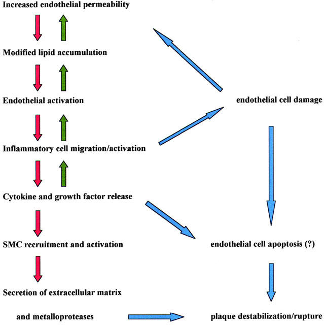
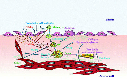
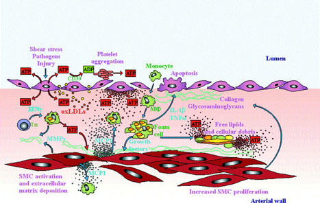
Atherosclerosis is a focal inflammatory disease of the arterial wall. It starts with the formation of fatty streaks on the arterial wall that evolve to form a raised plaque made of smooth muscle cells (SMCs), and infiltrating leukocytes surrounding a necrotic core. The pathogenesis of the atherosclerotic lesion is incompletely understood, but it is clear that a dysfunction of the endothelium, recruitment and activation of inflammatory cells and SMC proliferation have a pivotal role. Over recent years receptors for extracellular nucleotides, the P2 receptors, have been recognized as fundamental modulators of leukocytes, platelets, SMCs and endothelial cells. P2 receptors mediate chemotaxis, cytokine secretion, NO generation, platelet aggregation and cell proliferation in response to accumulation of nucleotides into the extracellular milieu. Clinical trials have shown the benefit of antagonists of the ADP platelet receptor(s) in the prevention of vascular accidents in patients with atherosclerosis. Therefore, we anticipate that a deeper understanding of the involvement of P2 receptors in atheroma formation will open new avenues for drug design and therapeutic intervention.
Figures
Similar articles
-
Leukotriene B4 signaling through NF-kappaB-dependent BLT1 receptors on vascular smooth muscle cells in atherosclerosis and intimal hyperplasia.Proc Natl Acad Sci U S A. 2005 Nov 29;102(48):17501-6. doi: 10.1073/pnas.0505845102. Epub 2005 Nov 17. Proc Natl Acad Sci U S A. 2005. PMID: 16293697 Free PMC article.
-
The role of inflammation in atherosclerosis.Isr J Med Sci. 1996 Nov;32(11):1059-65. Isr J Med Sci. 1996. PMID: 8960073 Review.
-
Role of redox signaling and poly (adenosine diphosphate-ribose) polymerase activation in vascular smooth muscle cell growth inhibition by nitric oxide and peroxynitrite.J Vasc Surg. 2008 Mar;47(3):599-607. doi: 10.1016/j.jvs.2007.11.006. J Vasc Surg. 2008. PMID: 18295111
-
New heterocyclic ligands for the adenosine receptors P1 and for the ATP receptors P2.Farmaco. 2005 Mar;60(3):185-202. doi: 10.1016/j.farmac.2004.09.007. Farmaco. 2005. PMID: 15784237
-
Involvement of P2 receptors in the growth and survival of neurons in the CNS.Pharmacol Ther. 2006 Mar;109(3):297-324. doi: 10.1016/j.pharmthera.2005.06.002. Epub 2005 Aug 15. Pharmacol Ther. 2006. PMID: 16102837 Review.
Cited by
-
CD39 and CD73 in immunity and inflammation.Trends Mol Med. 2013 Jun;19(6):355-67. doi: 10.1016/j.molmed.2013.03.005. Epub 2013 Apr 17. Trends Mol Med. 2013. PMID: 23601906 Free PMC article. Review.
-
Phosphorylation of beta-catenin by PKA promotes ATP-induced proliferation of vascular smooth muscle cells.Am J Physiol Cell Physiol. 2008 May;294(5):C1169-74. doi: 10.1152/ajpcell.00096.2008. Epub 2008 Mar 19. Am J Physiol Cell Physiol. 2008. PMID: 18353896 Free PMC article.
-
The adventitia: essential regulator of vascular wall structure and function.Annu Rev Physiol. 2013;75:23-47. doi: 10.1146/annurev-physiol-030212-183802. Epub 2012 Dec 3. Annu Rev Physiol. 2013. PMID: 23216413 Free PMC article. Review.
-
A Metabolomic Approach to Understanding the Metabolic Link between Obesity and Diabetes.Mol Cells. 2015 Jul;38(7):587-96. doi: 10.14348/molcells.2015.0126. Epub 2015 Jun 15. Mol Cells. 2015. PMID: 26072981 Free PMC article. Review.
-
Pharmacology of the new P2Y12 receptor inhibitors: insights on pharmacokinetic and pharmacodynamic properties.Drugs. 2013 Oct;73(15):1681-709. doi: 10.1007/s40265-013-0126-z. Drugs. 2013. PMID: 24114622 Review.
References
-
- BALAZOVICH K.J., BOXER L.A. Extracellular adenosine nucleotides stimulate protein kinase C activity and human neutrophil activation. J. Immunol. 1990;144:631–637. - PubMed
-
- BALSANO F., RIZZON P., VIOLI F., SCRUTINIO D., CIMMINIELLO C., AGUGLIA F., PASOTTI C., RUDELLI G. Antiplatelet treatment with ticlodipine in unstable angina. A controlled multicenter clinical trial. The Studio della Ticlodipina nell' Angina Instabile Group. Circulation. 1990;82:296–298. - PubMed
-
- BARICORDI O.R., FERRARI D., MELCHIORRI L., CHIOZZI P., HANAU S., CHIARI E., RUBINI M., DI VIRGILIO F. An ATP-activated channel is involved in mitogenic stimulation of human T lymphocytes. Blood. 1996;87:682–690. - PubMed
-
- BEIGI R.D., DUBYAK G.R. Endotoxin activation of macrophages does not induce ATP release and autocrine stimulation of P2 nucleotide receptors. J. Immunol. 2000;165:7189–7198. - PubMed
Publication types
MeSH terms
Substances
LinkOut - more resources
Full Text Sources
