. 2002 Mar;76(6):2654-66.
doi: 10.1128/jvi.76.6.2654-2666.2002.

Repression of African swine fever virus polyprotein pp220-encoding gene leads to the assembly of icosahedral core-less particles

Affiliations

Repression of African swine fever virus polyprotein pp220-encoding gene leads to the assembly of icosahedral core-less particles

Germán Andrés et al. J Virol. 2002 Mar.

Abstract

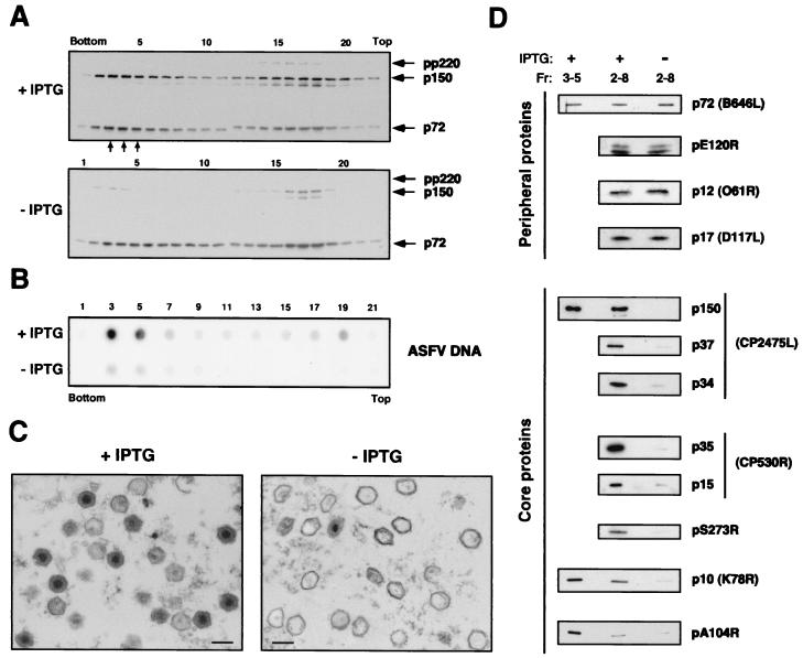
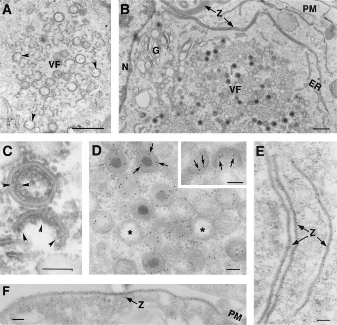
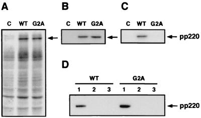
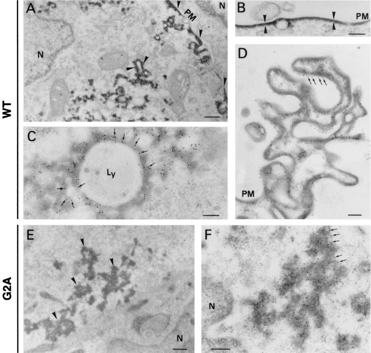
African swine fever virus (ASFV) polyprotein pp220, encoded by the CP2475L gene, is an N-myristoylated precursor polypeptide that, after proteolytic processing, gives rise to the major structural proteins p150, p37, p34, and p14. These proteins localize at the core shell, a matrix-like virus domain placed between the DNA-containing nucleoid and the inner envelope. In this study, we have examined the role of polyprotein pp220 in virus morphogenesis by means of an ASFV recombinant, v220i, containing an inducible copy of the CP2475L gene regulated by the Escherichia coli repressor-operator system. Under conditions that repress pp220 expression, the virus yield of v220i was about 2.6 log units lower than that of the parental virus or of the recombinant grown under permissive conditions. Electron microscopy revealed that pp220 repression leads to the assembly of icosahedral particles virtually devoid of the core structure. Analysis of recombinant v220i by immunoelectron microscopy, immunoblotting, and DNA hybridization showed that mutant particles essentially lack, besides the pp220-derived products, a number of major core proteins as well as the viral DNA. On the other hand, transient expression of the CP2475L gene in COS cells showed that polyprotein pp220 assembles into electron-dense membrane-bound coats, whereas a mutant nonmyristoylated version of pp220 does not associate with cellular membranes but forms large cytoplasmic aggregates. Together, these findings indicate that polyprotein pp220 is essential for the core assembly and suggest that its myristoyl moiety may function as a membrane-anchoring signal to bind the developing core shell to the inner viral envelope.


Figures

FIG. 1.
FIG. 1.
(A) Genomic structure of the ASFV recombinant virus v220i. The recombinant virus v220i was obtained from vGUSREP, a BA71V-derived recombinant virus, which contains the Lac repressor-encoding gene lacI inserted into the nonessential thymidine kinase locus. In the v220i virus, the promoter of the polyprotein pp220-encoding gene CP2475L was replaced by an inducible promoter, p72.I, which is composed of a strong late promoter (p72.4) and the operator sequence O1 (•) from the E. coli lac operon. The reporter genes lacZ and gusA, used for selection and purification of the recombinants, are also shown. (B) Plaque phenotype of v220i. Monolayers of Vero cells were infected with parental BA71V or recombinant v220i in the presence (+) or absence (−) of 0.15 mM IPTG. Plaques were visualized with 1% crystal violet 5 days after infection. (C) One-step growth curves of v220i. Vero cells were infected with 5 PFU of v220i per cell in the presence or absence of 0.15 mM IPTG. At the indicated times of infection, the total virus titer of each sample was determined by plaque assay on Vero cells in the presence of the inducer. Parental BA71V infections were also titrated as a control. Recombinant v220i was also grown under restrictive conditions for 12, 18, or 24 h and then induced with IPTG. At different times after induction, the infectious virus was titrated as described above.
FIG. 2.
FIG. 2.
Inducible expression of polyprotein pp220. Vero cells were either mock infected (lane M) or infected with parental BA71V or recombinant v220i virus in the presence (+) or absence (−) of IPTG. The cells were pulse-labeled with [35S]methionine-[35S]cysteine from 12 to 18 hpi, lysed, and either analyzed directly by SDS-PAGE (A) or first immunoprecipitated with a serum against polyprotein pp220 and its derived product p150 (B). The positions of the polyprotein pp220, the structural protein p150, the major capsid protein p72, and the reporter β-galactosidase and GUS proteins are indicated. Note that GUS protein migrates slightly slower than capsid protein p72. The electrophoretic mobilities of molecular mass markers are indicated at the left in panel A.
FIG. 3.
FIG. 3.
Electron microscopy of v220i-infected cells. Ultrathin Epon sections of v220i-infected cells incubated for 24 h in the presence (A, C, and F) or in the absence (B, D, E, and G) of IPTG are shown. While under permissive conditions (A), the assembly sites contain large amounts of mature virions, under restrictive conditions (B), they contain essentially icosahedral core-less particles. Compared with the intracellular mature virions (C), most of the defective particles (E) lack the core shell (cs) and the electron-dense nucleoid (n) but contain the inner envelope (ie) and the capsid (c). A minor population of mutant v220i particles (D) contain the nucleoid, but its size and position frequently appear to be altered. Defective v220i particles are released by budding at the plasma membrane (G), as occurs with the mature particles produced under permissive conditions (F). Bars, 200 nm (A, B, F, and G) and 100 nm (C, D, and E).
FIG. 4.
FIG. 4.
Immunoelectron microscopy of defective v220i particles. Vero cells were infected with recombinant v220i in the presence (A, C, E, and G) or absence (B, D, F, and H) of IPTG. At 24 hpi, the cells were fixed and processed by freeze-substitution. Ultrathin Lowicryl sections were incubated with antibodies against the capsid protein p72 (A and B), the core shell protein p150 (C and D), and the nucleoid protein pA104R (E and F), followed by incubation with protein A-gold (10 nm diameter). Sections were also labeled with a monoclonal anti-DNA antibody followed by a goat anti-mouse antibody conjugated to 10-nm-diameter gold particles (G and H). The arrowheads indicate representative labeling of the capsids (A and B), the core shells (C and D), and the nucleoids (E, F, G, and H) of particles at the virus factories. Note that p72 labeling is detected to similar extents on virions produced under permissive and nonpermissive conditions, while the labeling of core proteins and DNA is significantly weaker on the defective v220i particles. The images in panels D, F, and H have been selected to show one example of positively labeled virions (arrowheads). Bars, 100 nm.
FIG. 5.
FIG. 5.
Purification and analysis of extracellular defective v220i particles. (A) Extracellular virions collected from clarified culture supernatants of permissive (+ IPTG) and restrictive (− IPTG) v220i infections were adjusted to the same p72 content and subjected to equilibrium sedimentation in a Percoll gradient. Aliquots of the gradient fractions were analyzed by Western immunoblotting with antibodies against protein p72 and pp220-derived protein p150. The small arrows below fractions 3 to 5 of the + IPTG gradient indicate the position where the infectious ASFV particles band. The positions of proteins pp220, p150, and p72 are indicated. (B) DNA content of v220i particles. Alternate fractions of the Percoll gradients were analyzed by dot blot DNA hybridization with an ASFV probe. (C) Electron microscopy of purified v220i particles obtained in the presence or absence of IPTG. In both samples, fractions 2 to 8 of the Percoll gradients were pooled, gel filtrated through a Sephacryl S-1000 column, and processed by conventional Epon embedding. The material between virus particles mostly corresponds to clusters of Percoll particles. Bars, 200 nm. (D) Western immunoblotting of purified v220i particles with a spectrum of antibodies against ASFV structural proteins. Purified virus particles obtained under permissive (+) and nonpermissive (−) conditions were adjusted to the same p72 content and analyzed by SDS-PAGE and immunoblotting with antibodies to the indicated viral proteins. The mutant v220i particles (−) were collected from fractions 2 to 8 of the Percoll gradients, while the control virions (+) were recovered from fractions 2 to 8 or from fractions 3 to 5. Semipurified particles obtained from clarified culture media were used to analyze the proteinase pS273R.
FIG. 6.
FIG. 6.
Effect of pp220 induction on ASFV assembly. Electron microscopy of v220i-infected cells maintained for 12 h in the absence of IPTG (A) or induced from 12 to 24 hpi (B, C, D, E, and F). The samples were processed by conventional Epon embedding (A, B, and C) or by freeze-substitution followed by immunogold labeling with an antibody against pp220 and p150 (D, E, and F). Images in panels A to D correspond to virus factories, whereas those in panels E and F correspond to adjacent areas. Open particles are indicated by arrowheads in panel A. The arrowheads in panel C indicate the core shell of assembling particles. The arrows in panel D and in the insert indicate gold particles (10 nm) at the core shell of a mature virion and two open developing particles, respectively. The asterisks in panel D indicate core-less virions. Panels E and F show zipper-like structures associated with membranes from the ER or from the plasma membrane, respectively. N, nucleus; G, Golgi complex; PM, plasma membrane; VF, virus factories; Z, zipper-like structures. Bars, 500 nm (A and B) and 100 nm (C, D, E, and F).
FIG. 7.
FIG. 7.
Transient expression of polyprotein pp220 using the vaccinia virus-T7 RNA polymerase system. COS-7 cells were transfected with plasmid pGEM-T7 containing the wild-type (WT) CP2475L gene or a mutagenized copy (G2A) in which the N-terminal glycine codon was replaced by an alanine codon. pGEM-T7-transfected cells were used as a negative control (lanes C). After transfection, the cells were infected with vTF7-3, a vaccinia virus recombinant expressing T7 RNA polymerase, and labeled with [35S]methionine-[35S]cysteine or with [3H]myristic acid from 3 to 6 hpi. (A) SDS-PAGE of extracts of cells labeled with [35S]methionine-[35S]cysteine. (B) Immunoprecipitation of the 35S-labeled lysates with an anti-pp220 antibody. (C) Immunoprecipitation of the 3H-labeled extracts with an anti-pp220 antibody. (D) Cells transfected with the wild-type and mutagenized versions of the CP2475L gene were fractionated into a low-speed (500 × g) sediment (lanes 1) and a postnuclear supernatant, which was in turn fractionated into a soluble cytoplasmic fraction (lanes 2) and a high-speed (100,000 × g) sediment (lanes 3). The position of polyprotein pp220 is indicated.
FIG. 8.
FIG. 8.
Subcellular localization of myristoylated and nonmyristoylated pp220 in transfected COS-7 cells. Transfected COS-7 cells expressing myristoylable (wild-type [WT]) (A, B, C, and D) and nonmyristoylable (G2A) (E and F) polyprotein pp220 were processed by conventional Epon embedding (A and B) or by freeze-substitution and Lowicryl embedding (C, D, E, and F). The Lowicryl sections were incubated with an antibody against polyprotein pp220 followed by protein A-gold (10 nm diameter). Note that the myristoylated polyprotein assembles into electron-dense membrane coats (arrowheads in panels A and B), whereas the nonmyristoylated polyprotein forms large cytoplasmic aggregates (arrowheads in panel E). The arrowheads in panel B indicate the regular thickness (about 30 nm) of a dense coat bound to the plasma membrane. The arrows in panels C and D indicate pp220 labeling at dense coats bound to lysosomes and the plasma membrane, whereas the arrows in panel F indicate pp220 labeling at dense cytoplasmic aggregates. N, nucleus; PM, plasma membrane; Ly, lysosomes. Bars, 500 nm (A and E) and 200 nm (B, C, D, and F).

References

    1. Alcami, A., A. Angulo, C. Lopez-Otín, M. Muñoz, J. M. Freije, A. L. Carrascosa, and E. Viñuela. 1992. Amino acid sequence and structural properties of protein p12, an African swine fever virus attachment protein. J. Virol. 66:3860-3868. - PMC - PubMed
    1. Alves de Matos, A. P., and Z. G. Carvalho. 1993. African swine fever virus interaction with microtubules. Biol. Cell 78:229-234. - PubMed
    1. Andrés, G., C. Simón-Mateo, and E. Viñuela. 1997. Assembly of African swine fever virus: role of polyprotein pp220. J. Virol. 71:2331-2341. - PMC - PubMed
    1. Andrés, G., R. García-Escudero, C. Simón-Mateo, and E. Viñuela. 1998. African swine fever virus is enveloped by a two-membraned collapsed cisterna derived from the endoplasmic reticulum. J. Virol. 72:8988-9001. - PMC - PubMed
    1. Andrés, G., A. Alejo, C. Simón-Mateo, and M. L. Salas. 2001. African swine fever virus protease: a new viral member of the SUMO-1-specific protease family. J. Biol. Chem. 276:780-787. - PubMed

Publication types