. 2002 Mar;76(6):2721-9.
doi: 10.1128/jvi.76.6.2721-2729.2002.

Interaction between STAT-3 and HNF-3 leads to the activation of liver-specific hepatitis B virus enhancer 1 function

Affiliations

Interaction between STAT-3 and HNF-3 leads to the activation of liver-specific hepatitis B virus enhancer 1 function

Gulam Waris et al. J Virol. 2002 Mar.

Abstract

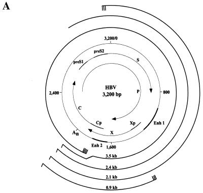
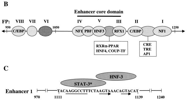
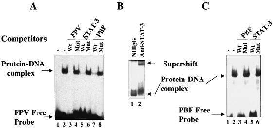
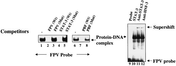
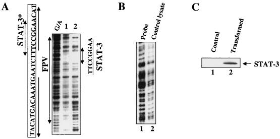
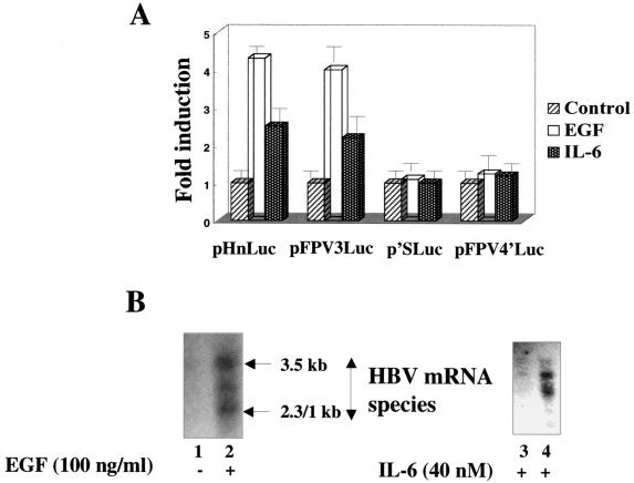
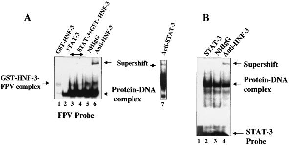
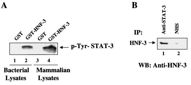
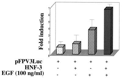
The signal transducer and activator of transcription 3 (STAT-3), a member of the STAT family of proteins, binds to a large number of transcriptional control elements and regulates gene expression in response to cytokines. While it binds to its cognate nucleotide sequences, it has been recently shown to directly interact with other transcriptional factors in the absence of DNA. We report here one such novel interaction between STAT-3 and hepatocyte nuclear factor 3 (HNF-3) in the absence of DNA. We have identified a STAT-3 binding site within the core domain of hepatitis B virus (HBV) enhancer 1. The HBV enhancer 1 DNA-STAT-3 protein interaction is shown to be stimulated by interleukin-6 (IL-6) and epidermal growth factor, which leads to an overall stimulation of HBV enhancer 1 function and viral gene expression. Using mobility shift assays and transient transfection schemes, we demonstrate a cooperative interaction between HNF-3 and STAT-3 in mediating the cytokine-mediated HBV enhancer function. Cytokine stimulation of HBV gene expression represents an important regulatory scheme of direct relevance to liver disease pathogenesis associated with HBV infection.


Figures

FIG. 1.
FIG. 1.
Schematic representations of the HBV genome and enhancer 1 element. (A) The viral genome is numbered (0 to 3,200 bp) according to the adw2 subtype of HBV. S, C, P, and X represent the viral genes encoding the surface antigen, core or e antigen, DNA polymerase, and X proteins, respectively. The HBV promoters (preS1, preS2, Cp, and Xp) and enhancers (Enh 1 and Enh 2) are indicated. An represents the single polyadenylation site used by all of the HBV RNAs. Each viral transcript is labeled with its respective length in kilobases (kb) and a poly(A) tail at the 3′ end. Multiple transcription initiation sites are indicated at the 5′ end of the 3.5-, 2.1-, and 0.9-kb RNAs. (B) Protein binding sites on Enh 1 are shown and footprint designations previously defined (38) are indicated above the corresponding sites. (C) The core domain spans nt 1080 to 1165 and is composed of the STAT-3 binding site, which is located at nt 1111 to 1127. The HNF-3 binding site spans nt 1124 to 1139. The DNA sequence of the STAT-3/HNF-3 region is shown with the corresponding cellular factors as indicated above. The thin line and arrows below the STAT-3-HNF-3 region indicate the positions of STAT-3 and FPV direct repeats, respectively.
FIG. 1.
FIG. 1.
Schematic representations of the HBV genome and enhancer 1 element. (A) The viral genome is numbered (0 to 3,200 bp) according to the adw2 subtype of HBV. S, C, P, and X represent the viral genes encoding the surface antigen, core or e antigen, DNA polymerase, and X proteins, respectively. The HBV promoters (preS1, preS2, Cp, and Xp) and enhancers (Enh 1 and Enh 2) are indicated. An represents the single polyadenylation site used by all of the HBV RNAs. Each viral transcript is labeled with its respective length in kilobases (kb) and a poly(A) tail at the 3′ end. Multiple transcription initiation sites are indicated at the 5′ end of the 3.5-, 2.1-, and 0.9-kb RNAs. (B) Protein binding sites on Enh 1 are shown and footprint designations previously defined (38) are indicated above the corresponding sites. (C) The core domain spans nt 1080 to 1165 and is composed of the STAT-3 binding site, which is located at nt 1111 to 1127. The HNF-3 binding site spans nt 1124 to 1139. The DNA sequence of the STAT-3/HNF-3 region is shown with the corresponding cellular factors as indicated above. The thin line and arrows below the STAT-3-HNF-3 region indicate the positions of STAT-3 and FPV direct repeats, respectively.
FIG. 2.
FIG. 2.
STAT-3 binds within HBV enhancer 1. (A) The EMSA was carried out in the presence of 32P-labeled FPV probe and STAT-3 protein synthesized in bacteria. Lane 1, probe alone; lane 2, 1 μg of STAT-3 protein; lanes 3, 5 , and 7 , 100-fold excesses of wild-type unlabeled FPV, STAT-3, and PBF oligonucleotides, respectively; lanes 4, 6, and 8, 100-fold excesses of mutant FPV, STAT-3, and PBF oligonucleotides, respectively. (B) The EMSA was carried out as described above but with the inclusion of normal human IgG (lane 1) and anti-STAT-3 serum (lane 2). (C) The EMSA was carried out in the presence of 32P-labeled PBF probe. Lane 1, probe alone; lane 2, 1 μg of STAT-3 protein; lanes 3 and 5, 100-fold excesses of unlabeled wild-type PBF and STAT-3 oligonucleotides, respectively; lanes 4 and 6, 100-fold excesses of mutant PBF and STAT-3 oligonucleotides, respectively.
FIG. 3.
FIG. 3.
Interaction between STAT-3 and FPV of HBV enhancer 1. The EMSA was carried out in the presence of 32P-labeled FPV probe and lysates from Huh-7 cells transfected with v-Src. Lanes 1 and 6, 5 μg of lysates as a source of STAT-3; lanes 2, 4, and 7, 100-fold excesses of wild-type FPV, STAT-3, and PBF oligonucleotides, respectively; lanes 3, 5, and 8, 100-fold excesses of mutant FPV, STAT-3, and PBF oligonucleotides, respectively; lanes 11 and 12, protein-DNA complex incubated with anti-STAT-3 and anti-HNF-3 serum, respectively.
FIG. 4.
FIG. 4.
DNase I protection analysis of STAT-3 binding. (A) Samples were analyzed in the presence of the enhancer 1 probe (25,000 cpm; labeled at nt 1308). Lanes 1 and 2 contained 50 and 100 μg of STAT-3 protein, respectively. G/A, sequencing ladder. The STAT-3 binding site is shown (nt 1111 to 1121) with a double-headed arrow indicating an 8-bp palindrome. ∗, previously designated as the PBF binding site (28). (B) DNase I protection assay. Lane 1, probe; lane 2, probe incubated with bacterial lysates lacking STAT-3 expression vector. (C) Western blot analysis. Bacterial lysates lacking STAT-3 vector (lane 1), doubly transformed with STAT-3 and JAK vectors (lane 2) were subjected to SDS-7.5% PAGE followed by immunoblot analysis using anti-STAT-3 polyclonal antibodies.
FIG. 5.
FIG. 5.
(A) Cytokines upregulate the STAT-3-dependent stimulation of HBV enhancer 1 in vivo . Huh-7 cells were transiently transfected with pHnLuc, pFPV3Luc, pFPV4′Luc, and p′SLuc reporter plasmids, followed by stimulation with EGF and IL-6 as described in Materials and Methods. The experiments were performed in duplicate. Luciferase activity is expressed as fold induction over the baseline level of activity observed during transfection of these vectors without EGF and IL-6 treatment. Each reporter plasmid is indicated below the graph. (B) Cytokine-mediated stimulation of HBV mRNA biosynthesis. Equal amounts of RNA from 2.2.1.5 cells either untreated or treated with EGF and IL-6 were analyzed by Northern blot analysis. RNA was transferred onto a nitrocellulose filter and probed with 32P-labeled 48-bp HBV DNA representing the X gene. Lane 1, RNA before stimulation was used as control; lane 2, incubation with EGF (6 h); lanes 3 and 4, stimulation with IL-6 for 6 and 12 h, respectively. Arrows indicate the 3.5- and 2.3- and 2.1-kb HBV mRNA species.
FIG. 6.
FIG. 6.
Cooperation of interaction between STAT-3 and HNF-3. (A) The EMSA was carried out in the presence of 32P-labeled FPV probe. Binding of FPV probe with recombinant GST-HNF-3 (lane 1); with STAT-3 synthesized in bacteria (lane 2); both STAT-3 and increasing concentration of HNF-3 (lanes 3 and 4); same as shown in lane 4 but incubated with normal human IgG (lane 5); or with anti-HNF-3 serum (lane 6) and with anti-STAT-3 serum (lane 7). (B) STAT-3-HNF-3 complex interacts with STAT-3 oligonucleotide probe. The EMSA was carried out in the presence of 32P-labeled STAT-3 oligonucleotide probe and nuclear lysates from HepG2 cells transfected with v-Src vector. Lane 1, probe alone; lane 2, 5 μg of lysates (source of STAT-3-HNF-3 complex); DNA-protein complex treated with normal human IgG (lane 3) or with anti-HNF-3 serum (lane 4).
FIG. 7.
FIG. 7.
(A) Direct interaction between STAT-3 and HNF-3. Nuclear extract from Huh-7 cells transfected with v-Src expression vector or STAT-3 synthesized in bacteria was incubated with GST (lanes 1 and 3) and GST-HNF-3 (lanes 2 and 4) bound to glutathione-Sepharose beads. STAT-3 bound to HNF-3 was extensively washed and subjected to SDS-7.5% PAGE followed by immunoblot analysis using anti-STAT-3 polyclonal antibodies. (B) In vivo interaction between STAT-3 and HNF-3. Huh-7 v-Src-transfected lysates were immunoprecipitated (IP) with anti-STAT-3 serum (lane 1) and normal human serum (NHS) (lane 2) fractionated by SDS-PAGE and electroblotted onto a PVDF membrane. The membrane was incubated with anti-HNF-3 serum followed by incubation with a secondary antibody and analyzed by ECL, a commercial detection kit.The arrow indicates the coimmunoprecipated HNF-3 in the STAT-3-HNF-3 in vivo complex.
FIG. 8.
FIG. 8.
Cooperative activation of HBV Enh 1 by STAT-3 and HNF-3. Huh-7 cells were transiently transfected with 1 μg each of pFPV3Luc reporter plasmid and HNF-3 vectors. At 36 h after transfection, the cells were serum starved overnight and then stimulated with EGF (100 ng/ml) for 15 min. The cells were harvested and analyzed for luciferase activity. Each transfection was carried out in duplicate and repeated at least three times.

Similar articles

Cited by

References

    1. Akira, S., Y. Nishio, M. Inoue, M. Wang, X. J. Wei, S. Matsusaka, T. Yoshida, T. Sudo, M. Naruto, and T. Kishimoto. 1994. Molecular cloning of APRF, a novel IFN-stimulated gene factor 3 p91-related transcription factor involved in the gp130-mediated signaling pathway. Cell 77:6371. - PubMed
    1. Antonucci, T. K., and W. J. Rutter. 1989. Hepatitis B virus (HBV) promoters are regulated by the HBV enhancer in a tissue-specific manner. J. Virol. 63:579-583. - PMC - PubMed
    1. Beasely, R. P., and L. Y. Hwang. 1984. Epidemiology of hepatocellular carcinoma. In G. N. Vyas et al. (ed.), Viral hepatitis and liver diseases. Grune & Stratton, New York, N.Y.
    1. Becker, S., B. Groner, and C. W. Muller. 1998. Three-dimensional structure of the Stat3β homodimer bound to DNA. Nature 349:145-151. - PubMed
    1. Bromberg, J. F., M. H. Wrzeszczynska, G. Devgan, Y. Zhao, R. G. Pestell, C. Albanese, and J. E. Darnell, Jr. 1999. Stat3 as an oncogene. Cell 98:295-303. - PubMed

Publication types

MeSH terms

LinkOut - more resources