

doi: 10.1128/jvi.76.6.3023-3030.2002.
Expression and self-assembly of norwalk virus capsid protein from venezuelan equine encephalitis virus replicons
Affiliations
- PMID: 11861868
- PMCID: PMC135954
- DOI: 10.1128/jvi.76.6.3023-3030.2002
Item in Clipboard
Expression and self-assembly of norwalk virus capsid protein from venezuelan equine encephalitis virus replicons
J Virol.
2002 Mar.
Abstract
The Norwalk virus (NV) capsid protein was expressed using Venezuelan equine encephalitis virus replicon particles (VRP-NV1). VRP-NV1 infection resulted in large numbers of recombinant NV-like particles that were primarily cell associated and were indistinguishable from NV particles produced from baculoviruses. Mutations located in the N-terminal and P1 domains of the NV capsid protein ablated capsid self-assembly in mammalian cells.
Figures

Expression of NLV capsid genes from VEE replicons. (A) Organization and transcription strategy of VEE replicons. In VEE replicons, the structural genes (capsid, E1, and PE2) are replaced with the NV capsid gene inserted into a polycloning site (PCS) under control of the subgenomic 26S promoter. Replicon transcripts coelectroporated into cells with the VEE defective packaging construct RNAs (VEE capsid and envelope glycoproteins) result in the packaging and release of infectious VEE VRPs that can be used as single-hit vaccine vectors in mammals (30, 41). Transfection will also result in the transient expression of high concentrations of rNV capsid protein which may self-assemble into NV VLPs. Upon infection of cells with VEE VRPs encoding the NV capsid gene, these single-hit replicon vectors will express high levels of the rNV capsid protein that self-assemble into ∼38-nm-diameter recombinant NLV particles which should be free of VEE VRPs. (B) Sequence of the NV major capsid gene. The NV capsid gene was cloned from stool samples of two human volunteers challenged with NV. The forward primer (NVCAPF1 [5′-GAT TTCCAGCAAGGTCATAC-3′]) and reverse primer (NVCAPR1 [5′-TTCGCCACCAACCTGTATGC-3′]) were designed to amplify the entire NV capsid region following reverse transcription-PCR (27, 46). The reaction was performed in a 50-μl reaction mixture with 5 μl of purified viral mRNA. Reverse transcription was performed at 50°C for 60 min, and then the temperature was elevated to 94°C for 2 min. Forty amplification cycles were performed, with one cycle consisting of 30 s at 94°C, annealing at 55°C for 1 min, and primer extension at 68°C for 2 min. While the NV1 sequence was identical to the published sequence (26, 27), the sequence of NV2 contained three amino acid changes. Chimeric constructs were assembled in which the N-terminal amino acid alterations in NV2 were joined in frame to the C terminus of NV1 (NV3) and in the opposite orientation (NV4). Using overlapping extension PCR, the NV capsid gene was fused to the VEE subgenomic promoter and inserted into the polycloning site of the VEE PVR21 plasmid vector and sequenced. (C) Expression of NV capsid proteins in cells from different mammals. Cells were infected with VRP-NV1 for 1 h at room temperature at a MOI of 5. At 18 h postinfection, the cultures were fixed and stained by FA staining for the presence of NV capsid proteins using NV antiserum from an infected volunteer using previously described techniques (5). Caco2 human colorectal adenocarcinoma cells, CRFK feline kidney cells, DBT murine astrocytoma cells, and swine testicular (ST) cells are shown.

Expression of NLV capsid genes from VEE replicons. (A) Organization and transcription strategy of VEE replicons. In VEE replicons, the structural genes (capsid, E1, and PE2) are replaced with the NV capsid gene inserted into a polycloning site (PCS) under control of the subgenomic 26S promoter. Replicon transcripts coelectroporated into cells with the VEE defective packaging construct RNAs (VEE capsid and envelope glycoproteins) result in the packaging and release of infectious VEE VRPs that can be used as single-hit vaccine vectors in mammals (30, 41). Transfection will also result in the transient expression of high concentrations of rNV capsid protein which may self-assemble into NV VLPs. Upon infection of cells with VEE VRPs encoding the NV capsid gene, these single-hit replicon vectors will express high levels of the rNV capsid protein that self-assemble into ∼38-nm-diameter recombinant NLV particles which should be free of VEE VRPs. (B) Sequence of the NV major capsid gene. The NV capsid gene was cloned from stool samples of two human volunteers challenged with NV. The forward primer (NVCAPF1 [5′-GAT TTCCAGCAAGGTCATAC-3′]) and reverse primer (NVCAPR1 [5′-TTCGCCACCAACCTGTATGC-3′]) were designed to amplify the entire NV capsid region following reverse transcription-PCR (27, 46). The reaction was performed in a 50-μl reaction mixture with 5 μl of purified viral mRNA. Reverse transcription was performed at 50°C for 60 min, and then the temperature was elevated to 94°C for 2 min. Forty amplification cycles were performed, with one cycle consisting of 30 s at 94°C, annealing at 55°C for 1 min, and primer extension at 68°C for 2 min. While the NV1 sequence was identical to the published sequence (26, 27), the sequence of NV2 contained three amino acid changes. Chimeric constructs were assembled in which the N-terminal amino acid alterations in NV2 were joined in frame to the C terminus of NV1 (NV3) and in the opposite orientation (NV4). Using overlapping extension PCR, the NV capsid gene was fused to the VEE subgenomic promoter and inserted into the polycloning site of the VEE PVR21 plasmid vector and sequenced. (C) Expression of NV capsid proteins in cells from different mammals. Cells were infected with VRP-NV1 for 1 h at room temperature at a MOI of 5. At 18 h postinfection, the cultures were fixed and stained by FA staining for the presence of NV capsid proteins using NV antiserum from an infected volunteer using previously described techniques (5). Caco2 human colorectal adenocarcinoma cells, CRFK feline kidney cells, DBT murine astrocytoma cells, and swine testicular (ST) cells are shown.

Expression of NLV capsid genes from VEE replicons. (A) Organization and transcription strategy of VEE replicons. In VEE replicons, the structural genes (capsid, E1, and PE2) are replaced with the NV capsid gene inserted into a polycloning site (PCS) under control of the subgenomic 26S promoter. Replicon transcripts coelectroporated into cells with the VEE defective packaging construct RNAs (VEE capsid and envelope glycoproteins) result in the packaging and release of infectious VEE VRPs that can be used as single-hit vaccine vectors in mammals (30, 41). Transfection will also result in the transient expression of high concentrations of rNV capsid protein which may self-assemble into NV VLPs. Upon infection of cells with VEE VRPs encoding the NV capsid gene, these single-hit replicon vectors will express high levels of the rNV capsid protein that self-assemble into ∼38-nm-diameter recombinant NLV particles which should be free of VEE VRPs. (B) Sequence of the NV major capsid gene. The NV capsid gene was cloned from stool samples of two human volunteers challenged with NV. The forward primer (NVCAPF1 [5′-GAT TTCCAGCAAGGTCATAC-3′]) and reverse primer (NVCAPR1 [5′-TTCGCCACCAACCTGTATGC-3′]) were designed to amplify the entire NV capsid region following reverse transcription-PCR (27, 46). The reaction was performed in a 50-μl reaction mixture with 5 μl of purified viral mRNA. Reverse transcription was performed at 50°C for 60 min, and then the temperature was elevated to 94°C for 2 min. Forty amplification cycles were performed, with one cycle consisting of 30 s at 94°C, annealing at 55°C for 1 min, and primer extension at 68°C for 2 min. While the NV1 sequence was identical to the published sequence (26, 27), the sequence of NV2 contained three amino acid changes. Chimeric constructs were assembled in which the N-terminal amino acid alterations in NV2 were joined in frame to the C terminus of NV1 (NV3) and in the opposite orientation (NV4). Using overlapping extension PCR, the NV capsid gene was fused to the VEE subgenomic promoter and inserted into the polycloning site of the VEE PVR21 plasmid vector and sequenced. (C) Expression of NV capsid proteins in cells from different mammals. Cells were infected with VRP-NV1 for 1 h at room temperature at a MOI of 5. At 18 h postinfection, the cultures were fixed and stained by FA staining for the presence of NV capsid proteins using NV antiserum from an infected volunteer using previously described techniques (5). Caco2 human colorectal adenocarcinoma cells, CRFK feline kidney cells, DBT murine astrocytoma cells, and swine testicular (ST) cells are shown.
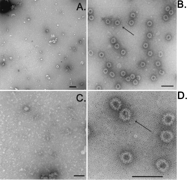
Assembly of NLV VLPs in mammalian cells. BHK cells (4 × 10⁷) were infected with VRP-NV1 or VRP-NV2. At 36 h postinfection, the cultures were freeze-thawed twice and putative particles were pelleted through a 20% sucrose cushion and resuspended in sterile phosphate-buffered saline. Viral suspensions were examined by negative staining with uranyl acetate (21). Grids were examined at 80 kV in a LEO EM-910 transmission electron microscope (Leo Electron Microscopy, Inc., Thornwood, N.Y.) at magnifications ranging from ×25,000 to ×125,000. Particles were visualized, photographed, and digitized prior to assembly of the images using Adobe Photoshop version 5.5 (Adobe Systems Inc.). VRP-NV2 (A and C) and VRP-NV1 (B and D) lysates at low and high magnifications are shown. Arrows indicate the rNV particles (VEE-Nor). Bars = 100 nm (B, C, and D) and 50 nm (A).

Cell association of recombinant NLV particles and mapping mutations which ablate particle formation. To determine if VEE-Nor particles are preferentially secreted from infected cells or remain in cellular compartments, BHK cells (4 × 10⁷ cells) were infected with VRP-NV1 at a MOI of 2 for 1 h. At 36 h postinfection, supernatants were harvested, cell monolayers were frozen twice, and cell debris was removed by centrifugation. Virus particles were concentrated from the supernatants or cell lysates by centrifugation through 20% sucrose cushions for 3 h at 28,000 rpm in a Beckman SW28 rotor and analyzed by electron microscopy as described in the legend to Fig. 2. Intracellular (A) and extracellular (B) VEE-Nor and particle formation from VEE-NV3- (C) and VEE-NV4- (D) transfected BHK cells (8.0 × 10⁶). Arrows indicate NLV particles. Bars = 100 nm.

VEE VRP expression of NV capsid proteins. BHK cells were infected with VRP-NV1 or VRP-NV2, and the cultures were harvested at 36 h postinfection. Cells were freeze-thawed three times, and the particles were purified by ultracentrifugation through 20% sucrose cushions or through 20 to 60% sucrose gradients. The purified particles were then pelleted by ultracentrifugation in an SW28 rotor. (A) Biochemical characterization of rNV capsid proteins synthesized in mammalian cells. Gradient-purified proteins were resuspended in Laemmli loading buffer, and 0.3 μg of purified protein was loaded per lane onto an SDS-10% polyacrylamide gel. After electrophoresis for 1 h at 180 V, the proteins were transferred to nitrocellulose, filters were blocked with 2% bovine serum albumin, and the immobilized proteins were probed following the standard Western blot protocol with a serum sample collected either predose or 21 days postdose from an NV-infected volunteer. Antibody-reactive protein was visualized with an alkaline phosphatase-conjugated anti-human immunoglobulin G secondary antibody (Sigma, St. Louis, Mo.) and 5-bromo-4-chloro-3-indolylphosphate (BCIP)-Nitro Blue Tetrazolium (NBT) substrate (Roche Molecular Biochemicals, Indianapolis, Ind.). The leftmost gel shows Coomassie blue staining of BAC-Nor and VRP-NV1 capsid proteins (Total rNV protein). Western blots using preimmune serum from a human volunteer prior to challenge with wild-type NV and postchallenge serum from the same human volunteer challenged with wild-type NV are shown. Arrows indicate the rNV proteins.

Posttranslational processing of rNV capsid proteins synthesized in mammalian cells. By Western blot analysis, VEE-Nor displayed a higher apparent molecular mass than that of BAC-Nor (rNV) (A). To remove potential conformational structure, VEE-Nor and BAC-Nor particles were further treated with 5 M guanidine-HCl at room temperature for 5 min to denature the proteins, the pH was adjusted to pH 8.6 with 4.6 M Tris, and the proteins were reduced with 0.1 M dithiothreitol (Sigma) for 45 min in the dark at room temperature before being alkylated by 0.25 M iodoacetic acid (Calbiochem) for 30 min at room temperature (23). The reduced and alkylated capsids were then diluted and analyzed by SDS-PAGE (B). (C) VEE-Nor was incubated with increasing concentrations (shown above the lanes) of endo-β-N-acetylglucosaminidase H (Endo H) (Roche Biochemicals) and peptide-N-glycosidase (Calbiochem) as previously described in the literature (38). Alkaline phosphatase (Alk Phos) (Roche Biochemicals) digestions were performed for 90 min at 37°C in 0.2 M Tris, pH 9.8. After enzymatic treatment, untreated capsid proteins were added to the reaction mixtures and the reaction mixtures were immediately boiled in loading buffer and separated on an SDS-polyacrylamide gel. A positive reaction is indicated by a doublet in a lane. Enzyme activity was verified using ovalbumin (ova) (Sigma) with each reaction set (data shown for Endo H treatment).
References
Publication types
MeSH terms
Grants and funding
LinkOut - more resources
Full Text Sources
Other Literature Sources
