. 2002 Mar 1;30(5):1154-62.
doi: 10.1093/nar/30.5.1154.

Phosphorylation of mammalian translation initiation factor 5 (eIF5) in vitro and in vivo

Affiliations

Phosphorylation of mammalian translation initiation factor 5 (eIF5) in vitro and in vivo

Romit Majumdar et al. Nucleic Acids Res. .

Abstract

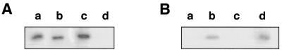
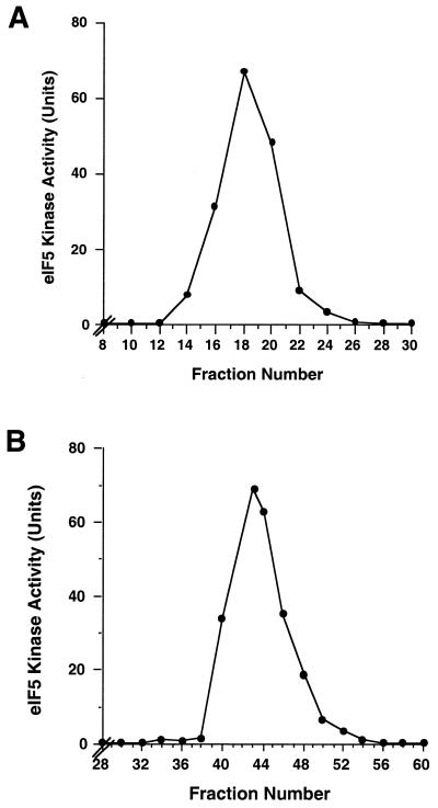
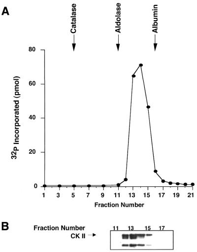
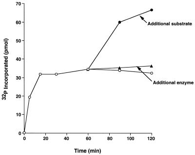
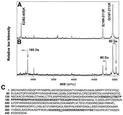
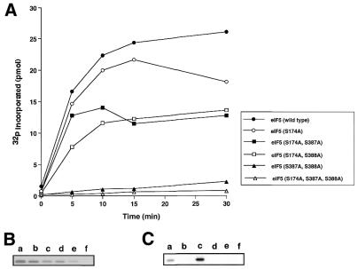
Eukaryotic translation initiation factor 5 (eIF5) interacts with the 40S initiation complex (40S*eIF3*AUG*Met-tRNA(f)*eIF2*GTP) and, acting as a GTPase activating protein, promotes the hydrolysis of bound GTP. We isolated a protein kinase from rabbit reticulocyte lysates on the basis of its ability to phosphorylate purified bacterially expressed recombinant rat eIF5. Physical, biochemical and antigenic properties of this kinase identify it as casein kinase II (CK II). Mass spectrometric analysis of maximally in vitro phosphorylated eIF5 localized the major phosphorylation sites at Ser-387 and Ser-388 near the C-terminus of eIF5. These serine residues are embedded within a cluster of acidic amino acid residues and account for nearly 90% of the total in vitro eIF5 phosphorylation. A minor phosphorylation site at Ser-174 was also observed. Alanine substitution mutagenesis at Ser-387 and Ser-388 of eIF5 abolishes phosphorylation by the purified kinase as well as by crude reticulocyte lysates. The same mutations also abolish phosphorylation of eIF5 when transfected into mammalian cells suggesting that CK II phosphorylates eIF5 at these two serine residues in vivo as well.


Figures

Figure 1
Figure 1
Phosphorylation of eIF5 by protein fractions derived from rabbit reticulocyte lysates. Purified, bacterially expressed eIF5 (5 pmol) was incubated with [γ-32P]ATP and the protein fractions derived from rabbit reticulocyte lysates in assay mixtures similar to that described in the Materials and Methods [Assay of eIF5-kinase activity (method A)]. The protein fractions used as the source of kinase were as follows. (A) Lanes: b, ribosomal 0.5 M KCl-wash protein fraction (15 µg); c, 0–50% ammonium sulfate fraction of post-ribosomal supernatant (16 µg); d, 50–80% ammonium sulfate fraction of post-ribosomal supernatant (76 µg). In lane a, eIF5 phosphorylated with 32P by recombinant CK II was used as a marker. (B) Phosphorylation of eIF5 with protein fractions obtained by chromatography of the 0–50% ammonium sulfate fraction of the post-ribosomal supernatant as described in the Materials and Methods (Purification of eIF5 kinase activity) and in the text. Lanes: a, flow-through fraction of first phosphocellulose chromatography (1.2 µg); b, 0.7 M phosphocellulose eluate (0.88 µg); c, DEAE-cellulose flow-through fraction of the phosphocellulose eluate (0.6 µg); d, 0.3 M DEAE-cellulose eluate (0.6 µg). Following incubation, the reaction mixtures were subjected to SDS–PAGE followed by autoradiography.
Figure 2
Figure 2
Elution of eIF5 kinase activity from phosphocellulose and FPLC-Mono Q columns. (A) Phosphocellulose chromatography of the 0.3 M DEAE-cellulose eluate. The pooled DEAE-cellulose 0.3 M eluate (11.3 mg protein), containing all the eIF5-kinase activity, was loaded onto a phosphocellulose column (12-ml bed volume) as described in the Materials and Methods (Purification of eIF5 kinase activity). Fractions of 3 ml were collected and assayed for eIF5 kinase activity using the assay method B as described in the Materials and Methods. (B) FPLC-Mono Q chromatography of the phosphocellulose protein fraction. The phosphocellulose eIF5 kinase fraction (150 U) was loaded onto a 1-ml bed volume of FPLC-Mono Q column as described in the Materials and Methods. Fractions of 0.5 ml were collected and assayed for eIF5 kinase activity.
Figure 3
Figure 3
Glycerol gradient centrifugation of eIF5 kinase activity. (A) The eIF5 kinase activity recovered from the FPLC-Mono Q column was concentrated to 400 µl by Centricon-30 filtration and layered onto a 10–30% (v/v) linear glycerol gradient (11 ml) as described in the Materials and Methods (Purification of eIF5 kinase activity). A marker gradient containing catalase, aldolase and albumin (2 mg each) was run in a parallel tube. The gradients were centrifuged in a SW41 rotor for 32 h at 40 000 r.p.m. at 4°C. Fractions (0.5 ml) were collected from the bottom of the tube and assayed for eIF5 kinase activity using the assay method B as described in the Materials and Methods. The sedimentation positions of the marker proteins are indicated. (B) The glycerol gradient fractions 11 through to 17 corresponding to eIF5 kinase activity (20 µl each) were analyzed by SDS–PAGE followed by immunoblotting with anti-CK II antibody.
Figure 4
Figure 4
Time course of eIF5 kinase activity. Reaction mixtures (20 ml) were prepared as described in the Materials and Methods except that 200 pmol of eIF5 and 0.5 µg of purified eIF5 kinase were added. Incubation was at 30°C. At the indicated times, aliquots (2 µl) were withdrawn and assayed for 32P incorporation into eIF5 using the assay method B. After 30 min of incubation, one series of tubes (indicated by ‘additional enzyme’) received an additional 0.5 µg of purified CK II, while another series (indicated by ‘additional substrate’) received an additional 200 pmol of eIF5 and incubation at 30°C was continued. At indicated times, 2 µl aliquots were withdrawn and assayed for 32P incorporation into eIF5 by assay method B. Open circles denote no further addition at 30 min, closed triangles denote additional 0.5 µg of CK II added at 30 min and closed circles denote additional 200 pmol of eIF5 added at 30 min. It should be noted that the results shown in the figure represent 32P incorporated into 2 µl reaction aliquot containing 20 pmol of eIF5.
Figure 5
Figure 5
MALDI-TOF mass spectra of tryptic peptides of unphosphorylated and phosphorylated eIF5. Unphosphorylated eIF5 (A) and phosphorylated eIF5 (B) were digested in solution with trypsin and pepsin and analyzed by MALDI-TOF without further separation. The tryptic peptides were labeled in (A) and their respective phosphorylated products were observed in (B) with mass differences of 80 or 160 Da. The vertical arrows represent the mass of the phosphorylated peptides while the horizontal arrows represent the mass shift from the unphosphorylated peptide. (C) The derived amino acid sequence of rat eIF5 taken from Si et al. (26). All the potential serine phosphorylation sites are shadowed. The serine residues at positions 174, 387 and 388 that are phosphorylated in vitro are underlined.
Figure 6
Figure 6
Analysis of eIF5 point mutations (Ser → Ala) for their ability to be phosphorylated in vitro. Reaction mixtures (80 µl each) containing 200 pmol of purified wild-type or mutant eIF5 proteins and 0.5 µg of purified eIF5 kinase were incubated at 30°C. At the indicated times, aliquots (5 µl) were withdrawn and assayed for 32P incorporation using either assay method B (A) or assay method A (B) as described in the Materials and Methods. It should be noted that the results shown in (A) represent 32P incorporated into 5 µl reaction aliquots containing 12.5 pmol of eIF5. (B) Lanes: a, wild-type eIF5; b, mutant eIF5 (S174A); c, mutant eIF5 (S174A, S387A); d, mutant eIF5 (S174A, S388A); e, mutant eIF5 (S387A, S388A); f, mutant eIF5 (S174A, S387A, S388A). (C) Wild-type or mutant (S174A, S387A, S388A) eIF5 proteins were incubated at 30°C with ribosomal 0.5 M KCl-wash fractions (lanes a and b, respectively) or with 0–50% ammonium sulfate fraction of the post-ribosomal supernatant (lanes c and d, respectively) or 50–80% ammonium sulfate fraction of the post-ribosomal supernatant (lanes e and f, respectively) under the conditions of eIF5 kinase assay as described in the Materials and Methods. After incubation at 30°C for 10 min, a 10 µl aliquot of each reaction mixture was subjected to SDS–PAGE followed by autoradiography of the dried gel.
Figure 7
Figure 7
Phosphorylation of eIF5 in mammalian cells. Human U2OS cells were transfected with eIF5 expression plasmids, pc DNA 3.1(+)Myc-HisB, containing either the wild-type eIF5 coding sequence or the mutant eIF5 coding sequence or with empty control vector. Following vector transfection, the cells were metabolically labeled with 32Pi as described in the Materials and Methods. eIF5 present in each cell lysate was immunoprecipitated with anti-Myc antibody and the washed immunocomplex was subjected to SDS–PAGE followed by electrophoretic transfer to a PVDF membrane. (A) The washed membrane blot was subjected to autoradiography. (B) The same membrane blot was subjected to immunoblot analysis using anti-Myc antibodies. The eIF5 expression plasmids used were as follows. Lanes: b, wild-type eIF5; c, mutant eIF5 (S387A, S388A); d, mutant eIF5 (S174A, S387A, S388A); e, control vector not containing the eIF5 coding sequence. In lane a, the wild-type, purified rat eIF5 protein (untagged), phosphorylated in vitro by purified CK II, was electrophoresed as a marker.
Figure 8
Figure 8
Effect of phosphorylation of eIF5 on its ability to form the 80S initiation complex. Reaction mixtures (50 µl) containing 20 mM Tris–HCl pH 7.5, 5 mM MgCl2, 100 mM KCl and 1 mM DTT (buffer R), 3 pmol of preformed [35S] Met-tRNAf•eIF2•GTP ternary complex (42 000 c.p.m./pmol), 1.2 A260 units of 40S ribosomal subunits and 0.1 A260 unit of the AUG codon were incubated for 5 min at 37°C to form the 40S initiation complex (40S•AUG•Met-tRNAf•eIF2•GTP). The chilled reaction mixtures were supplemented with 1.2 A260 units of 60S ribosomal subunits and 0.01 pmol of purified unphosphorylated or completely phosphorylated recombinant rat eIF5 as indicated. Following incubation at 37°C for 5 min, the chilled reaction mixtures were sedimented through a 5 ml linear 7.5–30% (w/v) sucrose density gradient in buffer R for 105 min at 48 000 r.p.m. at 4°C in a Beckman SW 50.1 rotor. Fractions (0.4 ml) collected from the bottom of each tube were counted for 35S radioactivity to quantify the formation of the 80S initiation complex.

Similar articles

Cited by

References

    1. Raychaudhuri P., Chaudhuri,A. and Maitra,U. (1985) Eukaryotic initiation factor 5 from calf liver is a single polypeptide chain protein of Mr = 62,000. J. Biol. Chem., 260, 2132–2139. - PubMed
    1. Chevesich J., Chaudhuri,J. and Maitra,U. (1993) Characterization of mammalian translation initiation factor 5 (eIF-5). Demonstration that eIF-5 is a phosphoprotein and is present in cells as a single molecular form of apparent Mr 58,000. J. Biol. Chem., 268, 20659–20667. - PubMed
    1. Das K., Chevesich,J. and Maitra,U. (1993) Molecular cloning and expression of cDNA for mammalian translation initiation factor 5. Proc. Natl Acad. Sci. USA, 90, 3058–3062. - PMC - PubMed
    1. Chakravarti D., Maiti,T. and Maitra,U. (1993) Isolation and immunochemical characterization of eukaryotic translation initiation factor 5 from Saccharomyces cerevisiae. J. Biol. Chem., 268, 5754–5762. - PubMed
    1. Chakravarti D. and Maitra,U. (1993) Eukaryotic translation initiation factor 5 from Saccharomyces cerevisiae. Cloning, characterization and expression of the gene encoding the 45,346-Da protein. J. Biol. Chem., 268, 10524–10533. - PubMed

Publication types