

Corkscrew retinal vessels in neurofibromatosis type 1: report of 12 cases
- PMID: 11864883
- PMCID: PMC1771041
- DOI: 10.1136/bjo.86.3.282
Corkscrew retinal vessels in neurofibromatosis type 1: report of 12 cases
Abstract
Aim: To describe a distinctive spectrum of retinal microvascular abnormalities in 12 patients with neurofibromatosis type 1 (NF-1).
Methods: This is an observational prospective study of the ocular fundus evaluated by direct ophthalmoscopy with or without fluorescein angiography, to investigate retinal microvascular abnormalities in 32 patients with NF-1 and in 30 control subjects. The evaluation included a complete general and neurological physical examination and in some cases computed tomography, magnetic resonance imaging with gadolinium-DTPA, or both.
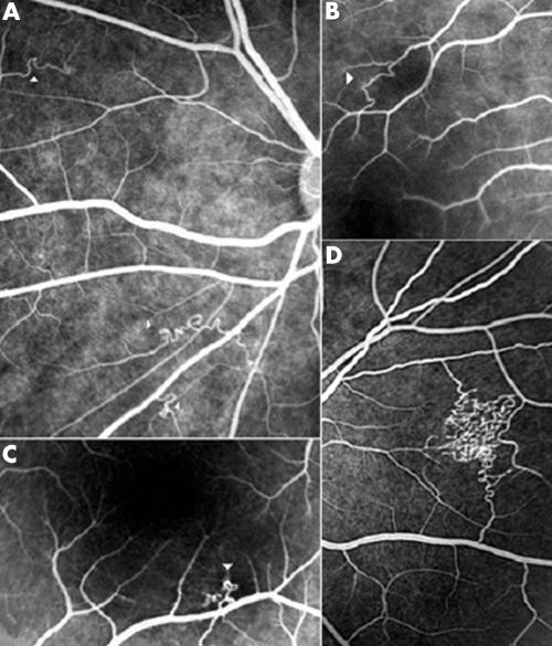
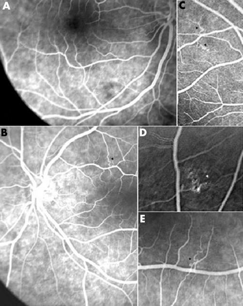
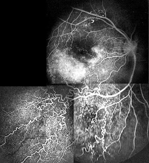
Results: The occurrence of a distinctive spectrum of retinal microvascular abnormalities is described in 12 patients with NF-1 (37.5%). At the lower end of the spectrum, present in 10 patients, the anomaly consisted of minuscule second or third order tortuous venules, which were called "corkscrew retinal vessels." These were usually isolated but in a few cases multiple. They flow towards the superior or inferior temporal veins. They had a length of one to two disc diameters. They ended either in a minute tuft or vanished on the retinal surface. The upper end of the spectrum was seen in only two patients. One of them had an exceptionally large venous anastomosis on the nasal retina and the other had an arteriovenous malformation extending over one retinal quadrant. None of the patients in the control group had such retinal microvascular abnormalities.
Conclusion: The "corkscrew" retinal vessels described in this report constitute a broad spectrum of microvascular markers in NF-1 patients.
Figures
References
-
- Destro M, D'Amico DJ, Gragoudas ES, et al. Retinal manifestations of neurofibromatosis. Diagnosis and management. Arch Ophthalmol 1991;109:662–6. - PubMed
-
- Yasunari T, Shiraki K, Hattori H, et al. Frequency of choroidal abnormalities in neurofibromatosis type 1. Lancet 2001;356:988–91. - PubMed
-
- National Institutes of Health Consensus Development Conference. Neurofibromatosis Conference Statement. Arch Neurol 1988;45:575–80. - PubMed
-
- Muci-Mendoza R. Anormalidades vasculares retinianas en neurofibromatosis con documentación de un hamartoma vascular puro no descrito. Gac Méd Caracas 1999;107:13–31.
MeSH terms
LinkOut - more resources
Full Text Sources
Research Materials
Miscellaneous