Case Reports
. 2002 Mar;86(3):321-7.
doi: 10.1136/bjo.86.3.321.

Polypoidal choroidal vasculopathy treated with macular translocation: clinical pathological correlation

Affiliations
Case Reports

Polypoidal choroidal vasculopathy treated with macular translocation: clinical pathological correlation

H Terasaki et al. Br J Ophthalmol. 2002 Mar.

Abstract

Aims: To report the histopathology of two specimens of polypoidal choroidal vasculopathy (PCV) obtained from two eyes of Japanese patients.

Methods: Specimens were obtained under direct visualisation during macular translocation surgery with 360 degree retinotomy. The clinical findings were correlated with the light microscopic findings of the two specimens.

Results: One specimen from a 77 year old man was the central portion of the lesion that lay under the sensory retina on the retinal pigment epithelium (RPE). The specimen was made up mainly of fibrous tissue with small, thin walled vessels. Indocyanine green angiography after surgery revealed that active leaking polypoidal element remained under the RPE. Another specimen obtained from a 62 year old man was made up of a fibrovascular membrane situated within Bruch's membrane. The part of this specimen inferior to the foveal region included a collection of dilated, thin walled blood vessels without pericytes, surrounded by macrophages that stained positive for CD68. The dilated vessels appeared to be correlated with the orange coloured polyps observed by ophthalmoscopy, the polypoidal structure seen in indocyanine green angiograms, and the pyramidal elevation with intermediate reflectivity by optical coherence tomography.

Conclusion: Polypoidal structures are located within Bruch's space. They are composed of clusters of dilated, thin walled blood vessels surrounded by macrophages and fibrin material. The positive immunohistochemical staining for vascular endothelial growth factor in the RPE and the vascular endothelial cells suggests that this fibrovascular complex is a subretinal choroidal neovascularisation.


Figures

Figure 1
Figure 1
Case 1. (A) Preoperative fundus photograph and vertical optical coherence tomographic scan (OCT) of the right eye. The yellow arrow points to the location of the cross sectional scan and possible pathological section. OCT shows an elevation of the sensory retina by a relatively highly reflective mass. The mass shows moderate internal reflection above the horizontal, linear, highly reflective band corresponding to the retinal pigment epithelium. (B) Fluorescein angiogram revealing a granular hyperfluorescence around the macula with a small amount of leakage around the fovea. (C) Indocyanine green angiogram demonstrating a branching vascular network superior to the fovea and polyp-like dilatations at the terminals of the branches nasosuperior to the fovea. (D) Postoperative fundus photograph and OCT at the newly located macula (white vertical bar = 250 μm). (E) Fluorescein angiogram 3 months after surgery showing the site of fluorescein leakage from the remaining polypoidal lesion at the previous macula. Also, cystoid macular oedema can be seen at the newly located macula. (F) Indocyanine green angiogram revealing polyp-like lesions with an associated network at the superior vascular arcade at the original site of the macula.
Figure 2
Figure 2
Case 2. (A) Preoperative fundus photograph and vertical optical coherence tomography scan (OCT) of the left eye. Macular oedema with lipid exudate, several orange-red lesions, and thin subretinal haemorrhage can be seen. OCT shows an oedematous elevation of the sensory retina. A small dome-shaped elevation showing another highly reflective line can be seen above the high reflective line corresponding to the retinal pigment epithelium. Two pyramidal elevations of the retinal pigment epithelium with moderate internal reflection at the upper and lower end of the vertical section corresponding to the orange-red lesions seen by ophthalmoscopy can be seen. (B) Fluorescein angiogram revealing granular hyperfluorescence around the macula with relative hypofluorescence at the centre. (C) Indocyanine green angiogram revealing subfoveal choroidal neovascularisation and polyp-like dilatations at the terminals of the branches around the fovea. The associated vascular networks cannot be seen. (D) Postoperative fundus photograph and OCT by vertical scan (white vertical bar = 250 μm.) (E) Fluorescein angiogram showing a large area of hyperfluorescence and extensive defect of the retinal pigment epithelium at the previous macula. An RPE rip can also be seen superior to the disc (arrow). (F) Indocyanine green angiogram showing atrophy of the choriocapillaris at the previous site of the macula without polypoidal lesions or vascular network.
Figure 3
Figure 3
Case 1, histopathology. (A) Subretinal fibrovascular tissue (haematoxylin and eosin, original magnification ×10). (B) A high power photomicrograph of the region outlined by the yellow box in the upper figure. The specimen is made up of mainly fibrous tissue with some small thin walled vessels (haematoxylin and eosin, original magnification ×100).
Figure 4
Figure 4
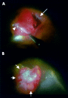
Case 2. Photographs from videophotography taken during surgery. (A) After peeling the thin fibrous membrane on the retinal pigment epithelium (short arrow), the subretinal pigment epithelial fibrovascular complex was removed with the retinal pigment epithelium (RPE). Large arrow indicates RPE tear. (B) Photograph of the choroidal side of the resected tissue under the operating microscope. White fibrous tissue is surrounded by clusters of relatively large vessels (arrows).
Figure 5
Figure 5
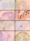
Case 2, histopathology. (A) Subretinal pigment epithelial fibrovascular tissue (haematoxylin and eosin, ×10). (B) A high power photomicrograph of the region outlined by the yellow box in (A). The fibrovascular tissue contains a collection of dilated, thin walled vessels that appear to correspond to the polypoidal lesions seen by ophthalmoscopy and indocyanine green angiography (haematoxylin and eosin, original magnification ×40). (C) Fibrovascular tissue can be seen under the basement membrane of the retinal pigment epithelium. (periodic acid Schiff, original magnification ×200). (D) The mass is seen under the elastic fibre layer of Bruch's membrane indicated by the arrows—that is, intra-Bruch's space (elastica van Gieson, original magnification ×400). Immunohistochemical and histochemical stains. (E) Abnormal vessels are lined by a thin endothelium without pericytes and are positive for CD34 (immunohistochemical stain for CD34, original magnification ×100). (F) Infiltration of macrophages around the large lumen vessels indicated by an arrow can be seen (immunohistochemical stain for the CD 68, original magnification ×100). (G) Massive leakage of fibrin material is seen in large area of specimen. Fibroblastic cells surrounded the fibrin materials around the vessels (phosphotungstic acid haematoxylin histochemical stain for fibrin, original magnification ×100). (H) Positive stain for antivascular endothelial growth factor in the retinal pigment epithelium and vascular endothelial cells (immunohistochemical stain for antivascular endothelial growth factor, original magnification ×100).

Similar articles

Cited by

References

    1. Yannuzzi LA. Idiopathic polypoidal choroidal vasculopathy. Presented at the 1982 Macula Society meeting.
    1. Yannuzzi LA, Sorenson J, Spaide RF, et al. Idiopathic polypoidal choroidal vasculopathy (IPCV). Retina 1990;10:1–8. - PubMed
    1. Stern RM, Zakov ZN, Zegarra H, et al. Multiple recurrent serosanguineous retinal pigment epithelial detachments in black women. Am J Ophthalmol 1985;100:560–9. - PubMed
    1. Perkovich BT, Zakov ZN, Berlin LA, et al. An update on multiple recurrent serosanguineous retinal pigment epithelial detachments in black women. Retina 1990;10:18–26. - PubMed
    1. Kleiner RC, Brucker AJ, Johnston RL. The posterior uveal bleeding syndrome. Retina 1990;10:9–17. - PubMed

Publication types