. 2002 Mar 4;156(5):817-28.
doi: 10.1083/jcb.200112059. Epub 2002 Feb 25.

14-3-3 transits to the nucleus and participates in dynamic nucleocytoplasmic transport

Affiliations

14-3-3 transits to the nucleus and participates in dynamic nucleocytoplasmic transport

Anne Brunet et al. J Cell Biol. .

Erratum in

  • J Cell Biol 2002 Apr 29;157(3):533

Abstract

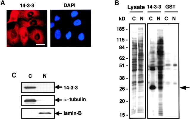
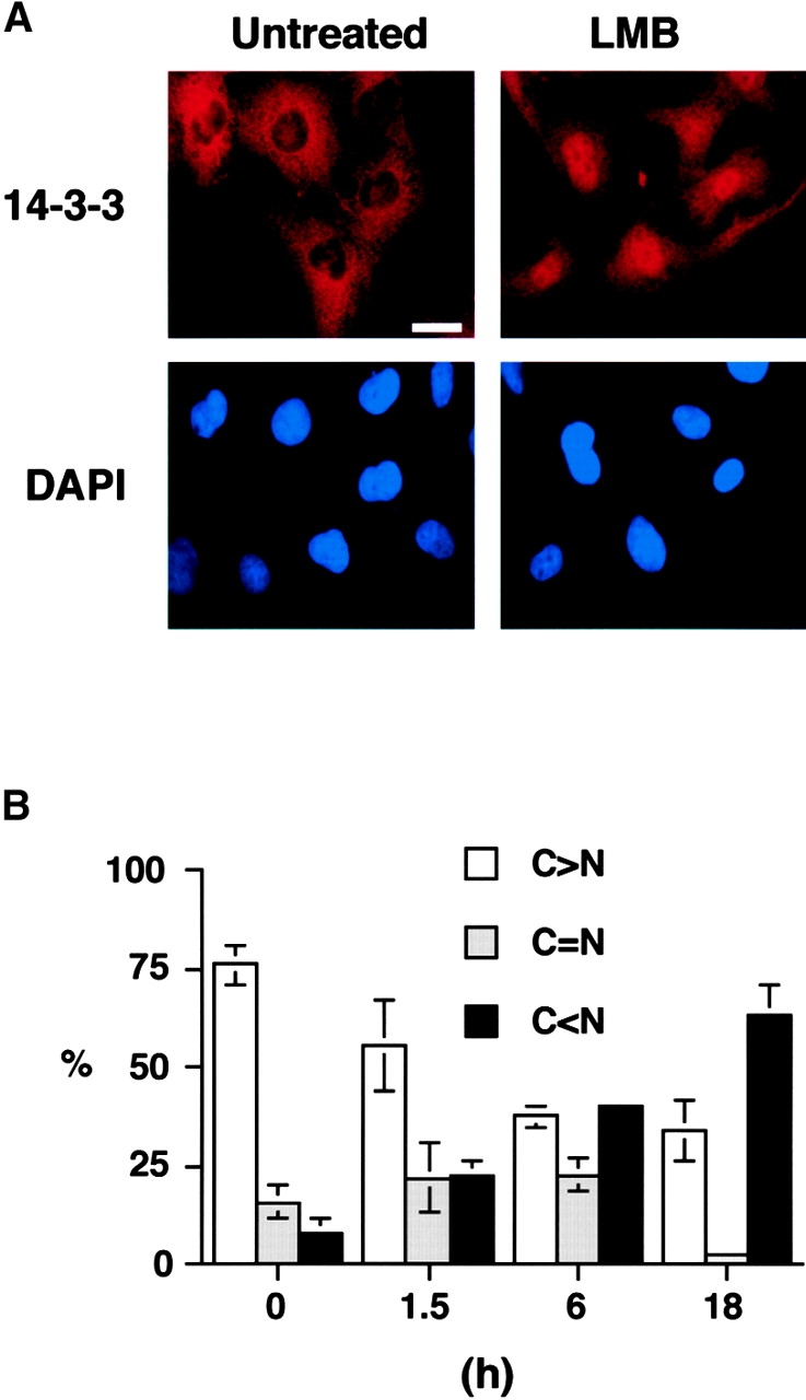
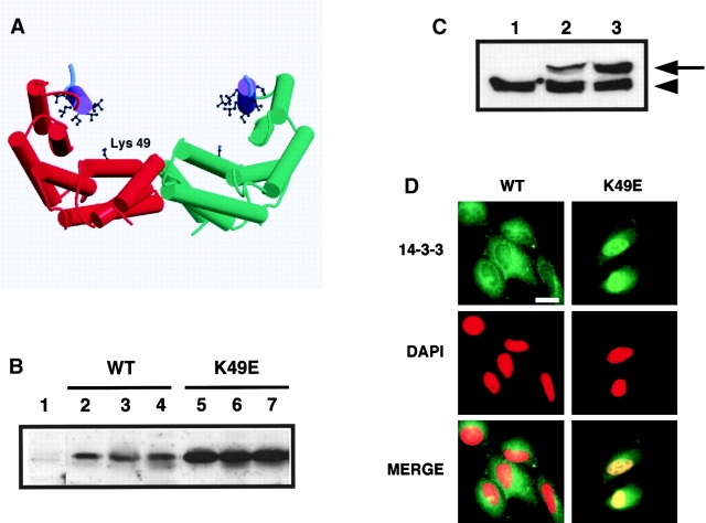
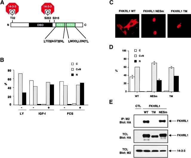
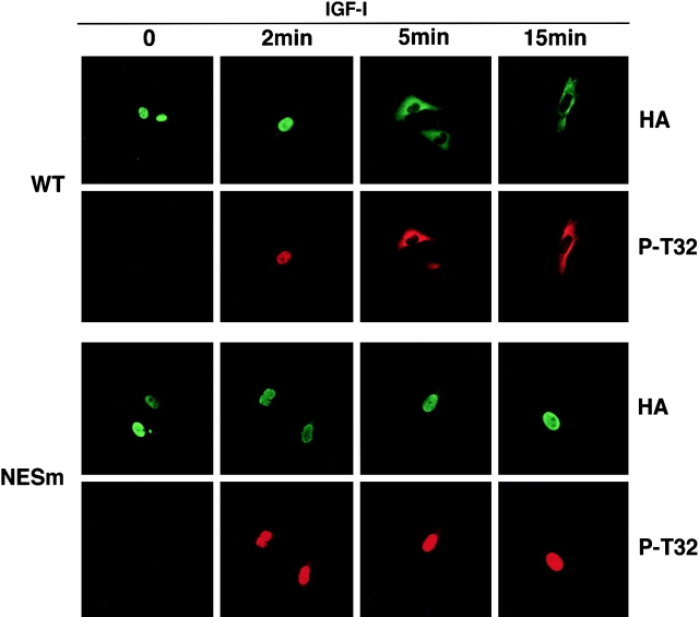
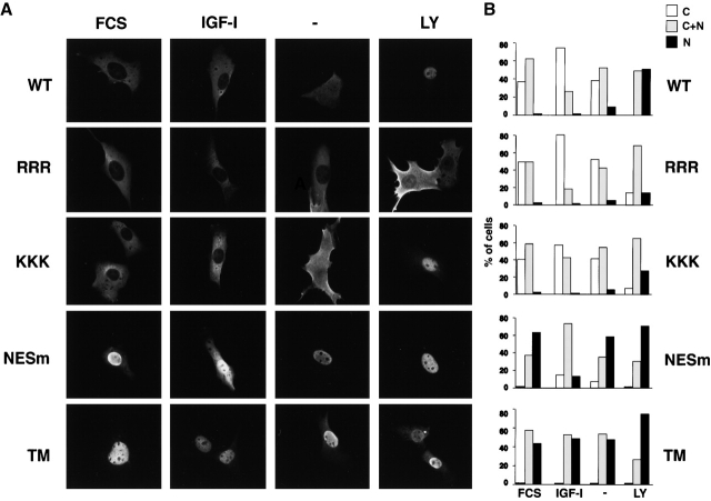
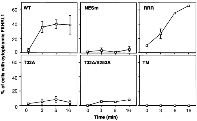
14-3-3 proteins regulate the cell cycle and prevent apoptosis by controlling the nuclear and cytoplasmic distribution of signaling molecules with which they interact. Although the majority of 14-3-3 molecules are present in the cytoplasm, we show here that in the absence of bound ligands 14-3-3 homes to the nucleus. We demonstrate that phosphorylation of one important 14-3-3 binding molecule, the transcription factor FKHRL1, at the 14-3-3 binding site occurs within the nucleus immediately before FKHRL1 relocalization to the cytoplasm. We show that the leucine-rich region within the COOH-terminal alpha-helix of 14-3-3, which had been proposed to function as a nuclear export signal (NES), instead functions globally in ligand binding and does not directly mediate nuclear transport. Efficient nuclear export of FKHRL1 requires both intrinsic NES sequences within FKHRL1 and phosphorylation/14-3-3 binding. Finally, we present evidence that phosphorylation/14-3-3 binding may also prevent FKHRL1 nuclear reimport. These results indicate that 14-3-3 can mediate the relocalization of nuclear ligands by several mechanisms that ensure complete sequestration of the bound 14-3-3 complex in the cytoplasm.


Figures

Figure 1.
Figure 1.
Different subcellular distributions of 14-3-3 and its ligands. (A) U2OS cells immunostained for 14-3-3σ (left) and corresponding DAPI staining (right). Bar, 10 μm. (B) Biochemical fractionation of U2OS cells followed by immunoblotting using an antibody recognizing all 14-3-3 isotypes (N, nuclear fraction; C, cytoplasmic fraction). Blots were probed with anti–α-tubulin (cytoplasmic) or anti–lamin B (nuclear) as controls. A very small amount of 14-3-3 is present in the nuclear fraction but cannot be seen in this exposure. (C) U2OS cells were metabolically labeled with [35S]methionine, fractionated, and nuclear (N) and cytoplasmic (C) fractions containing equal amounts of protein and radioactivity were incubated with beads containing GST–14-3-3 or GST alone. Bound proteins were visualized by SDS-PAGE and autoradiography. A small portion of the input sample is shown on the left (Lysate lanes). Positions of molecular weight markers, in kD, are indicated. The prominent band at ∼28 kD in the cytoplasmic/14-3-3–GST lane (arrow) is endogenous U2OS 14-3-3 (Rittinger et al., 1999).
Figure 2.
Figure 2.
LMB sequesters endogenous 14-3-3 in the nucleus. (A) Immunostaining of U2OS cells for endogenous 14-3-3σ with (right) or without (left) treatment with LMB for 18 h. Corresponding DAPI staining is shown on the bottom. Bar, 10 μm. (B) Quantitative analysis of 14-3-3σ location. Subcellular localization of 14-3-3σ was scored according to whether it was higher in the nucleus (N > C), evenly distributed between nucleus and cytoplasm (C = N), or higher in cytoplasm (C < N) for 300 cells at each time point. Results are the mean ± SD from three separate experiments.
Figure 3.
Figure 3.
The Leu-rich sequence in helix αI of 14-3-3 is responsible for ligand binding. (A) In the context of the entire 14-3-3 molecule, the Leu-rich helix αI functions primarily in ligand binding. Total cell lysates from 293T cells were incubated with beads containing the indicated GST–14-3-3 constructs or GST alone and bound proteins analyzed by SDS-PAGE and immunoblotting using an antibody against the 14-3-3 phosphoserine-binding epitope. TCL, total cell lysate; WT, WT 14-3-3; MT, K49E mutant; ΔC, GST–14-3-3 (1–214); ΔT, GST–14-3-3 (1–232). The mobility of molecular mass standards in kD is indicated. (B and C) Direct contribution of leucine and isoleucine residues in helix αI to ligand binding. 293T cell lysates as in A were incubated with beads containing immobilized WT GST–14-3-3ɛ or GST–14-3-3ɛ containing sequential Ala substitutions for Leu/Ile residues in helix αI. Bound proteins were analyzed by SDS-PAGE followed by immunoblotting using the antibody against the 14-3-3 phosphoserine-binding epitope (top) or stained with Coomassie blue to quantitate the amount of bead-bound GST–14-3-3 protein and GST alone (bottom, arrows). The bottom part of C shows the contributions from each Leu/Ile residue to phosphopeptide binding based on the x-ray structure of a 14-3-3–phosphopeptide complex (Rittinger et al., 1999).
Figure 4.
Figure 4.
A mutant 14-3-3, which cannot bind to ligands, accumulates in the nucleus. (A) Structural cartoon of the 14-3-3 dimer. Monomers (red and green) consists of nine α-helices. Helix αI containing the leucine-rich sequence is purple with leucine and isoleucine residues shown in ball and stick. Lys-49 in the phosphoserine-binding pocket is indicated. Only three residues in the extreme COOH-terminal tail (cyan) are visible in the x-ray structures. The figure was generated with Molscript and Raster3D. (B) Expression of Xpress-tagged 14-3-3 WT and K49E mutant protein in different stable U2OS cell lines. Total cell lysates corresponding to equivalent amounts of total protein from stable cell lines expressing empty vector (lane 1), WT 14-3-3 (lanes 2–4), or K49E mutant (lanes 5–7) were analyzed by SDS-PAGE and immunoblotting using an anti-Xpress epitope antibody. (C) Comparison of Xpress-tagged and endogenous 14-3-3 levels in several U2OS stable cell lines. Total cell lysates from stable cell lines containing empty vector (lane 1), WT 14-3-3ɛ (clone #15) (lane 2) and 14-3-3ɛ K49E mutant (clone #21) (lane 3) were immunoblotted using a pan anti–14-3-3 antibody. Arrowhead indicates endogenous 14-3-3 in U2OS cells; arrow indicates Xpress-tagged 14-3-3. (D) Immunostaining of stable U2OS cell lines expressing WT 14-3-3ɛ (clone #15, left) or 14-3-3ɛ K49E mutant (clone #21, right) with the anti-Xpress antibody. Corresponding DAPI staining was shown in the middle, and the merged image is shown on the bottom. Bar, 10 μm. Each of three stable cell lines showed identical results.
Figure 5.
Figure 5.
Both FKHRL1 nuclear export sequences and interaction with 14-3-3 are necessary for efficient nucleocytoplasmic transport. (A) Schematic illustration of FKHRL1 showing the T32, S253, and S315 phosphorylation sites and the 14-3-3 binding sites. The two NES sequences in FKHRL1 are shown (NES1, amino acids 369–378; NES2, amino acids 386–396). (B) LMB treatment prevents FKHRL1 nuclear export even in the presence of growth factors. CCL39 cells were transfected with a WT HA-FKHRL1 construct. Cells were starved for 20 h, incubated with LMB for 2 h, and then stimulated with 10% serum (FCS) or 100 ng/ml IGF-I for 15 min. Localization of FKHRL1 WT or mutants was monitored by immunolocalization with the anti-HA antibody. Quantitative analysis of a representative experiment is shown. C, cytoplasm; C + N, cytoplasm and nucleus; N, nucleus. (C) Mutation of FKHRL1 NES or 14-3-3 binding site prevents FKHRL1 relocalization from the nucleus to the cytoplasm in the presence of growth factors. CCL39 cells were transfected with a WT HA-FKHRL1 construct or a mutant of both NES mutant of FKHRL1 (NESm) or a mutant in which all three phosphorylation sites of FKHRL1 have been replaced by alanine (TM). Cells were incubated in the presence of 10% serum (FCS). Localization of FKHRL1 WT or mutants was monitored by immunolocalization with the anti-HA antibody. (D) Quantitative analysis of FKHRL1 localization. Results are the mean ± SD from three separate experiments. C, cytoplasm; C + N, cytoplasm and nucleus; N, nucleus. (E) Mutation of FKHRL1 NES does not abolish binding to 14-3-3, whereas mutation of the phosphorylation sites of FKHRL1 does. 293T cells were cotransfected with a construct encoding M2–14-3-3ζ and either empty vector (CTL) or vectors encoding HA-FKHRL1 WT, T32A/S253A/S315A (TM), or a mutant of the putative NES1 and 2 (NESm). 14-3-3 was immunoprecipitated with the anti-M2 antibody, and the immune complex was analyzed by SDS-PAGE and immunoblotted with the anti-HA antibody (top). Total cell lysates (TCL) were also analyzed by direct immunoblotting with the anti-HA antibody (middle) or the anti-M2 antibody (bottom).
Figure 6.
Figure 6.
Phosphorylation of FKHRL1 at the 14-3-3 binding site occurs within the nucleus immediately before nuclear export. CCL39 fibroblasts were transfected with WT HA-tagged FKHRL1 or a mutant of NES1 and -2 (NESm). Cells were serum starved for 12 h and incubated in the presence of 20 μM LY294002 for the last hour and then incubated in the presence of 100 ng/ml IGF-I for the indicated periods of time. Cells were rapidly fixed with paraformaldehyde, and FKHRL1 was detected by immunolocalization with an anti-HA antibody. Phosphorylation of FKHRL1 was assessed by immunolocalization with an antibody to phospho-Thr32. Representative pictures are shown. NESm, mutant of NES1 and NES2 of FKHRL1.
Figure 7.
Figure 7.
Role of NLS and NES sequences in FKHRL1 subcellular localization. (A) CCL39 fibroblasts were transfected with a WT HA-tagged FKHRL1 construct or with FKHRL1 mutant constructs. Cells were either incubated in the presence of 10% serum (FCS) or 100 ng/ml IGF-I (IGF-I), or serum-starved for 12 h (−), or incubated in the presence of 20 μM LY294002 for 1 h (LY). FKHRL1 was detected by immunolocalization with an anti-HA antibody. RRR, RRR → AAA mutant of FKHRL1; KKK, KKK → AAA mutant of FKHRL1; NESm, mutant of NES1 and NES2 of FKHRL1; TM, triple mutant of FKHRL1 (T32A/S253A/S315A). (B) Quantification of a representative experiment is shown.
Figure 8.
Figure 8.
Kinetics of FKHRL1 cytoplasmic relocalization after IGF-I stimulation shows a requirement for both 14-3-3 binding and NES sequences for rapid nuclear export and NLS for inhibition of nuclear reimport. CCL39 fibroblasts were transfected with a WT HA-tagged FKHRL1 construct or with various FKHRL1 mutant constructs. Cells were treated as in the legend to Fig. 6, and FKHRL1 was detected by immunolocalization using an anti-HA antibody. The data presented correspond to the mean and variation of two experiments. Error bars are present for all time points, though in some cases they are obscured by the plot symbol. RRR, RRR → AAA mutant of FKHRL1; NESm, mutant of NES1 and NES2 of FKHRL1; TM, triple mutant of FKHRL1 (T32A/S253A/S315A).

References

    1. Beck, T., and M.N. Hall. 1999. The TOR signalling pathway controls nuclear localization of nutrient-regulated transcription factors. Nature. 402:689–692. - PubMed
    1. Brownawell, A.M., G.J. Kops, I.G. Macara, and B.M. Burgering. 2001. Inhibition of nuclear import by protein kinase B (Akt) regulates the subcellular distribution and activity of the forkhead transcription factor AFX. Mol. Cell. Biol. 21:3534–3546. - PMC - PubMed
    1. Brunet, A., A. Bonni, M.J. Zigmond, M.Z. Lin, P. Juo, L.S. Hu, M.J. Anderson, K.C. Arden, J. Blenis, and M.E. Greenberg. 1999. Akt promotes cell survival by phosphorylating and inhibiting a forkhead transcription factor. Cell. 96:857–868. - PubMed
    1. Cahill, C.M., G. Tzivion, N. Nasrin, S. Ogg, J. Dore, G. Ruvkun, and M. Alexander-Bridges. 2001. Phosphatidylinositol 3-kinase signaling inhibits DAF-16 DNA binding and function via 14-3-3-dependent and 14-3-3-independent pathways. J. Biol. Chem. 276:13402–13410. - PubMed
    1. Dalal, S.N., C.M. Schweitzer, J. Gan, and J.A. DeCaprio. 1999. Cytoplasmic localization of human cdc25C during interphase requires an intact 14-3-3 binding site. Mol. Cell. Biol. 19:4465–4479. - PMC - PubMed

Publication types

MeSH terms