

Suprabasal p53 immunoexpression is strongly associated with high grade dysplasia and risk for malignant transformation in potentially malignant oral lesions from Northern Ireland
- PMID: 11865002
- PMCID: PMC1769595
- DOI: 10.1136/jcp.55.2.98
Suprabasal p53 immunoexpression is strongly associated with high grade dysplasia and risk for malignant transformation in potentially malignant oral lesions from Northern Ireland
Abstract
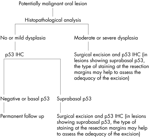
Aims: No good predictive marker for the malignant transformation of potentially malignant oral lesions (PMOLs) is currently available. This study re-evaluated the value of p53 immunoexpression to predict malignant transformation of PMOLs after discounting possible confounding factors.
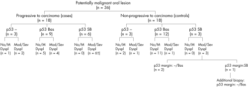
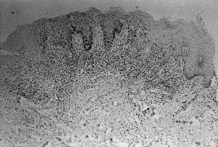
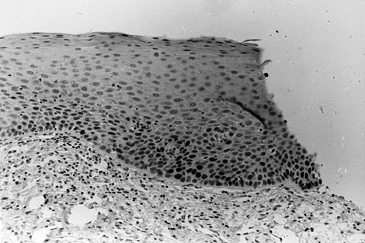
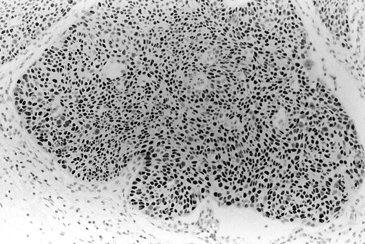
Methods: PMOLs from 18 patients who showed progression to carcinoma, 16 of the respective carcinomas, and PMOLs from 18 matched controls were evaluated by immunohistochemistry (IHC) for p53 expression. A mouse monoclonal antibody that detects wild-type and mutant forms of human p53 was used. The p53 immunostaining pattern was also correlated with the degree of dysplasia.
Results: Suprabasal p53 staining was significantly associated with high grades of dysplasia (p < 0.01). The specificity and positive predictive value (PPV) for malignant transformation of suprabasal p53 staining were superior to the assessment of dysplasia, but sensitivity was inferior. All carcinomas derived from PMOLs with suprabasal p53 showed strong p53 immunostaining. However, the absence of suprabasal p53 staining and/or dysplastic changes did not preclude malignant transformation in a considerable proportion of PMOLs.
Conclusions: This study confirms and extends previous findings that suprabasal p53 immunoexpression has a high PPV for malignant transformation of PMOLs and can be used as a specific marker for lesions that are at high risk for malignant transformation. The absence of suprabasal p53 staining (that is, absence of, or basal, p53 staining) is non-informative for prognostic purposes. Because of its limited sensitivity, p53 IHC is not a substitute for the assessment of dysplasia in the evaluation of PMOLs. Instead, p53 IHC emerges as a clinically useful supplement of histopathological assessment in the prognosis of PMOLs.
Figures
References
-
- Axéll T, Pindborgg JJ, Smith CJ, et al. Oral white lesions with special reference to precancerous and tobacco-related lesions: conclusions of an international symposium held in Uppsala, Sweden, May 18–21, 1994. J Oral Pathol Med 1996;25:49–54. - PubMed
-
- Leonardelli GB, Talamazzi F. Leucoplasie del caro orale e precancerosi, Nota I. Archivo Italiano di Otologia Rinologia e Laringologia 1950;61:107–14. - PubMed
-
- Schepman KP, van der Meij EH, Smeele LE, et al. Malignant transformation of oral leukoplakia: a follow-up study of a hospital-based population of 166 patients with oral leukoplakia from The Netherlands. Oral Oncol 1998;34:270–5. - PubMed
-
- Silverman S, Gorsky M, Lozada F. Oral leukoplakia and malignant transformation. A follow-up study of 257 patients. Cancer 1984;53:563–8. - PubMed
-
- Hansen LS, Olson JA, Silverman S, Jr. Proliferative verrucous leukoplakia: a long-term study of 30 patients. Oral Surg Oral Med Oral Pathol 1985;60:285–98. - PubMed
MeSH terms
Substances
LinkOut - more resources
Full Text Sources
Medical
Research Materials
Miscellaneous