. 2002 Mar;22(6):1664-73.
doi: 10.1128/MCB.22.6.1664-1673.2002.

RNA destabilization by the granulocyte colony-stimulating factor stem-loop destabilizing element involves a single stem-loop that promotes deadenylation

Affiliations

RNA destabilization by the granulocyte colony-stimulating factor stem-loop destabilizing element involves a single stem-loop that promotes deadenylation

R A Putland et al. Mol Cell Biol. 2002 Mar.

Abstract

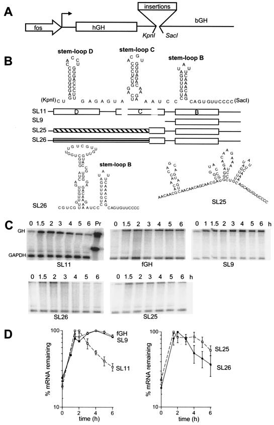
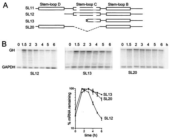
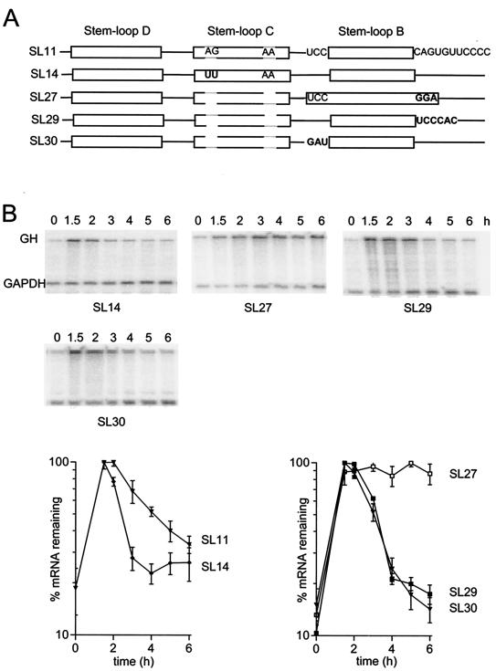
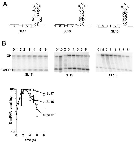
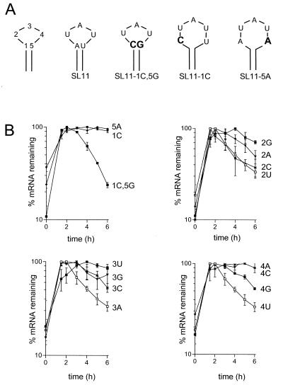
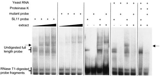
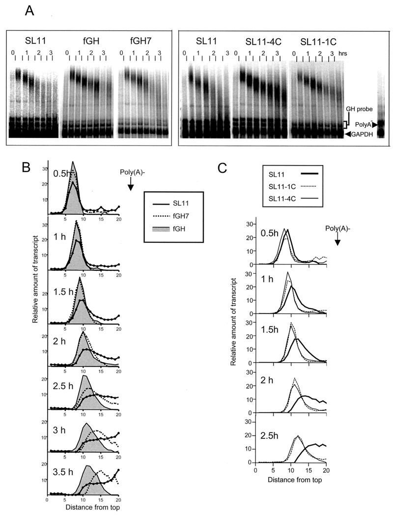
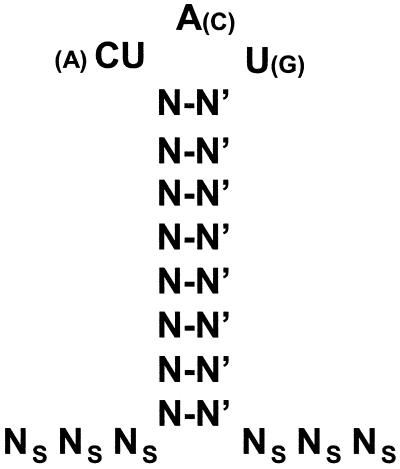
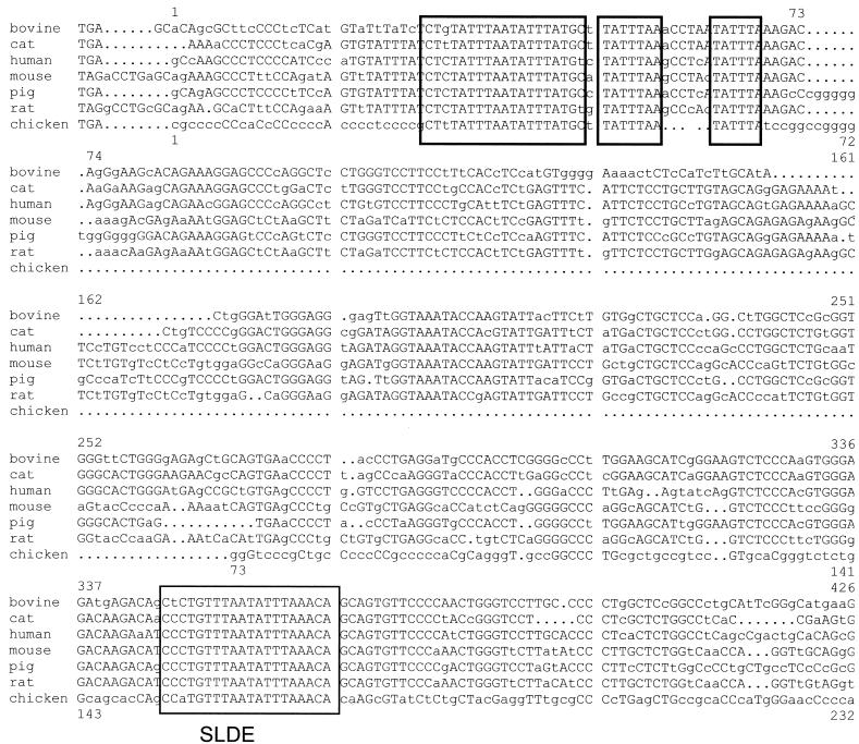
Granulocyte colony-stimulating factor (G-CSF) mRNA contains two distinct types of cis-acting mRNA destabilizing elements in the 3'-untranslated region. In addition to several copies of the AU-rich element the G-CSF mRNA also contains a destabilizing region that includes several predicted stem-loop structures. We report here that the destabilizing activity resides in a single stem-loop structure within this region. A consensus sequence for the active structure has been derived by site-directed mutagenesis, revealing that a three-base loop of sequence YAU and unpaired bases either side of the stem contribute to the activity. The helical nature of the stem is essential and the stem must be less than 11 bp in length, but the destabilizing activity is relatively insensitive to the sequence within the helix. The stem-loop increases the rate of mRNA deadenylation, most likely by enhancing the processivity of the deadenylation reaction. A protein that binds the stem-loop, but not an inactive mutant form, has been detected in cytoplasmic lysates.


Figures

FIG. 1.
FIG. 1.
Predicted secondary structure of a region from the 3′-UTR of G-CSF that contains a potent destabilizing element and the effect of deletion or replacement of some stem-loops. (A) Schematic of the fGH reporter gene. The transcription start site is indicated by an arrow. The translated region, which is derived from human growth hormone (hGH), is boxed. (B) The predicted structure of the SLDE region of SL11 and schematics showing the region deleted or replaced in SL9, SL25, and SL26. Stem-loops in the schematic diagram are shown as open boxes, while the regions replaced in SL25 and SL26 are shown as hatched boxes. The predicted structure in SL26 is also shown. (C) RNase protection assays. RNA was isolated at the indicated times after serum stimulation of NIH 3T3 cells stably transfected with variants of the fGH gene containing the indicated sequences inserted in the 3′-UTR. The upper band is the protection product from probe to the growth hormone region of the fGH reporter; the lower band is the protection product from the GAPDH internal control. The SL11 gel also shows the migration of undigested probes. (D) Time courses of the RNase protection data. Each line on the graphs is labeled with the sequence name. The data shown are means and standard errors of the means from four (SL11), three (fGH), or two (SL25, SL26) experiments.
FIG.2.
FIG.2.
The effect of deletion of individual stem-loops. (A) Schematic showing the truncated regions inserted into the fGH reporter. (B) RNase protection assays. RNA was isolated at the indicated times after serum stimulation of NIH 3T3 cells stably transfected with variants of the fGH gene containing the indicated sequences inserted in the 3′-UTR. The upper band is the protection product from probe for the growth hormone region of the fGH reporter; the lower band is the protection product from the GAPDH internal control. Representative RNase protection gels are shown for each construct and pooled data from two or more experiments are plotted below.
FIG.3.
FIG.3.
Lengthening stem-loop C enhances instability but lengthening stem-loop B abrogates destabilization. (A) Schematic showing the mutations introduced into SL11. (B) RNase protection assays. RNA was isolated at the indicated times after serum stimulation of NIH 3T3 cells stably transfected with variants of the fGH gene containing the indicated sequences inserted in the 3′-UTR. The upper band is the protection product from probe for the growth hormone region of the fGH reporter; the lower band is the protection product from the GAPDH internal control. Representative RNase protection gels are shown for each construct and pooled data from two or more experiments are plotted below.
FIG.4.
FIG.4.
The destabilizing effect requires base pairing in the stem but is not highly dependent on the sequence. (A) Schematic showing changes introduced into the stem of stem-loop B. Changed bases are shown in bold. Asterisks indicate the GC base pair that replaces the AU base pair that is present in the wild-type G-CSF. This substitution was previously shown to maintain destabilizing activity (5). (B) RNase protection assay gels and quantification. RNA was isolated at the indicated times after serum stimulation of NIH 3T3 cells stably transfected with variants of the fGH gene containing the indicated sequences inserted in the 3′-UTR. The upper band is the protection product from probe for the growth hormone region of the fGH reporter; the lower band is the protection product from the GAPDH internal control. Representative RNase protection gels are shown for each construct and pooled data from two or more experiments are plotted below.
FIG.5.
FIG.5.
The structure and sequence of the B loop are important for its function as a destabilizing element. (A) Schematic showing changes to the closing base pair of stem-loop B. The structure at left shows the numbering of bases used in naming of constructs. Changed bases are shown in bold. (B) RNA degradation time courses. Variant forms of SL11 with changes either to the closing base pair of stem-loop B as shown in panel A or point mutations in the three-base loop as indicated by the label on the time course were transfected and analyzed by RNase protection assay as described in Materials and Methods and the legend for Fig. 1.
FIG.6.
FIG.6.
Electrophoretic mobility shift detection of a complex on the SL11 RNA. SL11 probe corresponding to the sequence shown in Fig. 1 or a mutant probe containing a single inactivating point mutation in the SLDE loop (Fig. 5, SL11-4C) was incubated with cytoplasmic extract from BALB/c 3T3 cells and subjected to digestion with T1 nuclease, and the products were electrophoresed on a native 6% polyacrylamide gel. The first pair of panels show complexes resulting from incubation of SL11 and SL11-4C probes, respectively, with 0.625, 1.25, 2.5 and 5 μg of cytoplasmic protein extract. A complex that forms on the SLDE but not the mutant probe is indicated with arrows. Other panels show the complexes formed on SL11 or SL11-4C probe with or without 1 μg of protein extract, in the presence or absence of 1.6 μg of yeast RNA per ml plus 20 μg of tRNA per ml, and with or without digestion for 5 min by 10 μg of proteinase K per ml.
FIG. 7.
FIG. 7.
The SLDE enhances deadenylation. (A) To monitor poly(A) tail lengths, RNase A-resistant hybrids formed between the mRNA and a probe that spans the 3′ end of the mRNA were electrophoresed on nondenaturing gels. RNA was isolated at half-hour intervals after serum stimulation of NIH 3T3 cells stably transfected with SL11, fGH (a stable mRNA that deadenylates slowly), or fGH7 (which contains an ARE that enhances the deadenylation rate) (left panel) or with SL11 or two stable mutants of SL11 as shown (right panel). A GAPDH coding region probe was included as an internal standard. The lane on the right shows the migration from in vitro-prepared poly(A) RNA. Note that the two bands immediately above the GAPDH band are residual degradation products from the GH probe and are not due to accumulation of deadenylated mRNA in vivo. (B) To compare deadenylation rates, individual lanes from the left panel were scanned by phosphorimager and the signal intensity from the top of the gel to near the expected location of deadenylated RNA was plotted for each time point. The SL11 trace is shown as a solid line, the fGH7 trace is shown as a dotted line, and the fGH trace is shaded. (C) Plots of individual lanes from the right panel were scanned by phosphorimager and the signal intensity was plotted. The SL11 trace is shown as a thick line, the SL11-4C trace is shown as a thin line, and the SL11-1C trace is shown as a dashed line.
FIG. 8.
FIG. 8.
Consensus sequence for activity of the stem-loop destabilizing element. A schematic representation of the functional element summarizing the results of the mutagenesis data are shown. N-N′, an essential base pair whose sequence can vary; Ns, bases that must be single stranded. Optimal bases in the loop are shown in large print and suboptimal bases are shown in parentheses.
FIG. 9.
FIG. 9.
Comparison of part of the 3′-UTR sequences of G-CSF mRNAs from different species. Sequences were aligned using the GCG programs CLUSTAL and GAP. Regions conserved across all seven species are boxed. The lower box includes the SLDE. The sequences (and their GenBank accession numbers) are from cow (AF092533 [15]), cat (AB042552 [27]), human (X03438 [23]), mouse (M13926 [25]), pig (Y10494 [17]), rat (U37101 [13]), and chicken (X14477 [19]). The numbering shown above the sequences is with respect to the start of the human 3′-UTR and below is with respect to the start of the chicken 3′-UTR.

References

    1. Binder, R., J. A. Horowitz, J. P. Basilion, D. M. Koeller, R. D. Klausner, and J. B. Harford. 1994. Evidence that the pathway of transferrin receptor mRNA degradation involves an endonucleolytic cleavage within the 3′ UTR and does not involve poly(A) tail shortening. EMBO J. 13:1969-1980. - PMC - PubMed
    1. Binder, R., S.-P. L. Hwang, R. Ratnasabapathy, and D. L. Williams. 1989. Degradation of apolipoprotein II mRNA occurs via endonucleolytic cleavage at 5′-AAU-3′/5/-UAA-3′ elements in single-stranded loop domains of the 3′-noncoding region. J. Biol. Chem. 264:16910-16918. - PubMed
    1. Brewer, G., and J. Ross. 1988. Poly(A) shortening and degradation of the 3′ A+U-rich sequences of human c -myc mRNA in a cell-free system. Mol. Cell. Biol. 8:1697-1708. - PMC - PubMed
    1. Brown, B. D., I. D. Zipkin, and R. M. Harland. 1993. Sequence-specific endonucleolytic cleavage and protection of mRNA in Xenopus and Drosophila. Genes Dev. 7:1620-1631. - PubMed
    1. Brown, C. Y., C. A. Lagnado, and G. J. Goodall. 1996. A cytokine mRNA-destabilizing element that is structurally and functionally distinct from A+U-rich elements. Proc. Natl. Acad. Sci. USA 93:13721-13725. - PMC - PubMed

Publication types

MeSH terms

LinkOut - more resources