. 2002 Mar;22(6):1674-83.
doi: 10.1128/MCB.22.6.1674-1683.2002.

Caspase cleavage of initiation factor 4E-binding protein 1 yields a dominant inhibitor of cap-dependent translation and reveals a novel regulatory motif

Affiliations

Caspase cleavage of initiation factor 4E-binding protein 1 yields a dominant inhibitor of cap-dependent translation and reveals a novel regulatory motif

Andrew R Tee et al. Mol Cell Biol. 2002 Mar.

Abstract

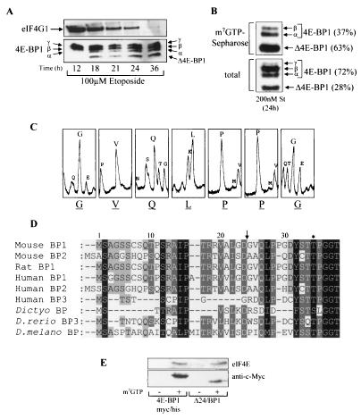
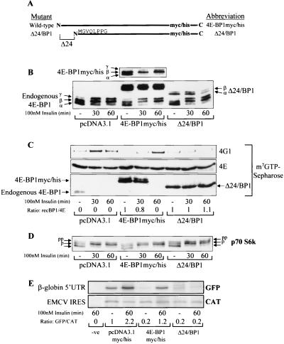
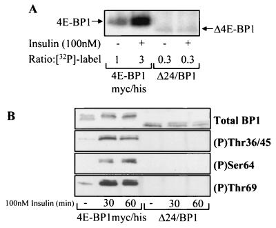
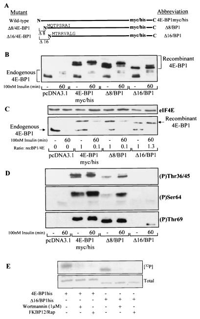
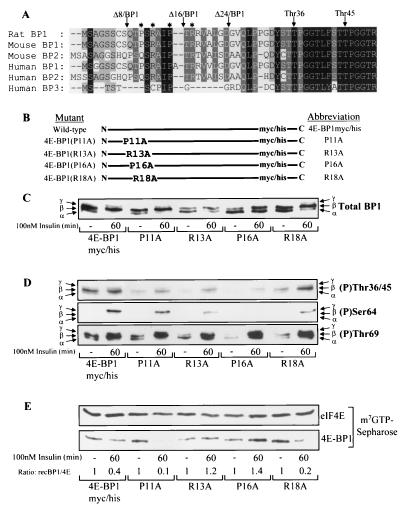
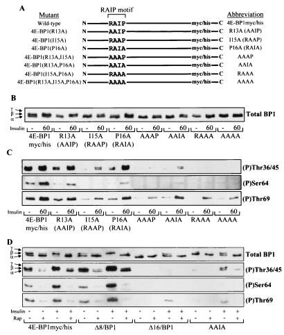
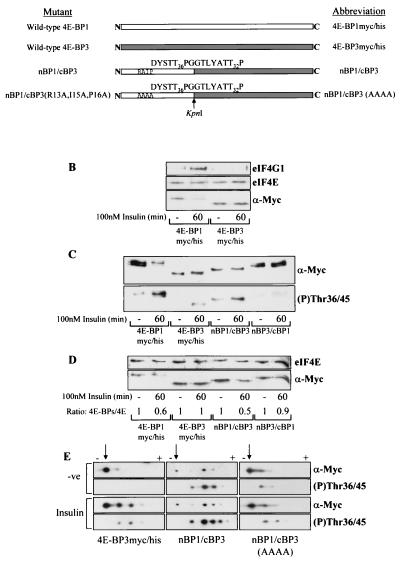
Eukaryotic initiation factor 4E (eIF4E) binding proteins (4E-BPs) regulate the assembly of initiation complexes required for cap-dependent mRNA translation. 4E-BP1 undergoes insulin-stimulated phosphorylation, resulting in its release from eIF4E, allowing initiation complex assembly. 4E-BP1 undergoes caspase-dependent cleavage in cells undergoing apoptosis. Here we show that cleavage occurs after Asp24, giving rise to the N-terminally truncated polypeptide Delta4E-BP1, which possesses the eIF4E-binding site and all the known phosphorylation sites. Delta4E-BP1 binds to eIF4E and fails to become sufficiently phosphorylated upon insulin stimulation to bring about its release from eIF4E. Therefore, Delta4E-BP1 acts as a potent inhibitor of cap-dependent translation. Using a mutagenesis approach, we identify a novel regulatory motif of four amino acids (RAIP) which lies within the first 24 residues of 4E-BP1 and which is necessary for efficient phosphorylation of 4E-BP1. This motif is conserved among sequences of 4E-BP1 and 4E-BP2 but is absent from 4E-BP3. Insulin increased the phosphorylation of 4E-BP3 but not sufficiently to cause its release from eIF4E. However, a chimeric protein that was generated by replacing the N terminus of 4E-BP3 with the N-terminal sequence of 4E-BP1 (containing this RAIP motif) underwent a higher degree of phosphorylation and was released from eIF4E. This suggests that the N-terminal sequence of 4E-BP1 is required for optimal regulation of 4E-BPs by insulin.


Figures

FIG. 1.
FIG. 1.
Identification of the caspase-dependent cleavage of 4E-BP1. (A) Swiss 3T3 cells were treated with etoposide for the times indicated. The samples were analyzed for total eIF4G1 (top) and 4E-BP1 (bottom). The α-, β-, and γ-species of 4E-BP1 and cleavage product Δ4E-BP1 are labeled. (B) Swiss 3T3 cells were treated with staurosporine (St) for 24 h, and extracts were subjected to affinity chromatography on m7GTP-Sepharose (see Materials and Methods). The percentages of uncleaved 4E-BP1 (γ, β, and α) and cleaved (Δ4E-BP1) 4E-BP1 bound to eIF4E (top) and total levels present within cell extracts (bottom) were quantified by densitometry (National Institutes of Health Image, version 1.61). (C) Δ4E-BP1 (isolated as described for panel B) was excised from the membrane and subjected to Edman degradation, yielding N-terminal sequence GVQLPPG. (D) N-terminal alignment of 4E-BPs. Arrow, cleavage site; ∗, phosphorylation site at Thr36 in mouse and rat 4E-BP1 and at Thr37 in human 4E-BP1. 4E-BP1 homologues from D. discoideum (Dictyo), D. rerio, and D. melanogaster (D. melano) are also shown. (E) Cell lysates prepared from serum-starved HEK293 cells overexpressing either 4E-BP1myc/his or Δ24/BP1 (see Fig. 2A for details of these mutants) were subjected to affinity chromatography using either m7GTP-Sepharose (+) or Sepharose (CL-4B; −). The levels of purified recombinant 4E-BP1 and eIF4E were analyzed.
FIG. 2.
FIG. 2.
The cleaved fragment of 4E-BP1 (Δ24/BP1) acts as a dominant inhibitor of eIF4F complex assembly and cap-dependent translation. (A) Diagrammatic representation of mutants 4E-BP1myc/his and Δ24/BP1. The amino acid sequence indicates the N-terminus of Δ24/BP1. (B) HEK293 cells were transiently transfected with the empty vector or pcDNA3.1myc/his expressing either 4E-BP1myc/his or Δ24/BP1, as indicated. These cells were serum starved and treated for 30 or 60 min (as indicated) with insulin. Cell extracts were analyzed to assess 4E-BP1 phosphorylation. 4E-BP1, endogenous 4E-BP1 phosphorylation; 4E-BP1myc/his (top), shorter exposure of the overexpressed 4E-BP1myc/his. (C) The cell extracts from panel B were subjected to affinity chromatography on m7GTP-Sepharose (see Materials and Methods). The amounts of eIF4G1 (4G1), eIF4E (4E), endogenous 4E-BP1 (4E-BP1), 4E-BP1myc/his, and Δ24/BP1 were analyzed. The ratios of recombinant 4E-BP1 bound to eIF4E (recBP1/4E) were assessed by densitometry (National Institutes of Health [NIH] Image, version 1.61). (D) p70 S6 kinase phosphorylation was analyzed. Arrows, differently phosphorylated species (pp, most-phosphorylated species; lowest arrow, least-phosphorylated species). (E) A bicistronic vector containing both a GFP reporter gene downstream from the β-globin 5′ untranslated region (UTR) and a CAT reporter gene downstream from the encephalomyocarditis virus (EMCV) IRES was cotransfected into HEK293 cells as indicated. Lane −ve, no-DNA control. Cell extracts were analyzed for cap-dependent translation as described in Materials and Methods. The ratios of the amounts of [35S]methionine incorporated into GFP and CAT were determined by densitometry (NIH Image, version 1.61); these ratios were normalized against the empty-vector serum-starved control (value set at 1).
FIG. 3.
FIG. 3.
The cleaved form of 4E-BP1 does not undergo efficient insulin-stimulated phosphorylation. (A) HEK293 cells overexpressing either 4E-BP1myc/his or Δ24/BP1. In vivo radiolabeling was carried out as described in Materials and Methods and 32P label incorporation into 4E-BP1 was quantified ([32P]-label; the 4E-BP1myc/his serum-starved-only sample is standardized to 1). (B) HEK293 cells overexpressing either 4E-BP1myc/his or Δ24/BP1 at comparable levels were treated as indicated. The extracts were analyzed for total 4E-BP1 levels and phosphorylation of 4E-BP1 at Thr36 and 45, Ser64, and Thr69 as indicated.
FIG. 4.
FIG. 4.
Residues between 8 and 16 are required for efficient phosphorylation of 4E-BP1. (A) Diagrammatic representation of mutants 4E-BP1myc/his, Δ8/BP1, and Δ16/BP1. The amino acid sequences indicate the N termini of these truncated proteins. (B) HEK293 cells were transiently transfected with plasmids expressing either 4E-BP1myc/his, Δ8/BP1, or Δ16/BP1 as indicated. The pcDNA3.1myc/his vector was used as a control. Cells were serum starved and treated with insulin as indicated. The extracts were analyzed by using the anti-4E-BP1 antibody. (C) The cell extracts from panel B were subjected to affinity chromatography on m7GTP-Sepharose as described for Fig. 2C. The ratios of recombinant 4E-BP1 bound to eIF4E (recBP1/4E) were assessed by densitometry (National Institutes of Health Image, version 1.61), where the ratio of recombinant 4E-BP1 to eIF4E under serum-starved conditions is normalized to unity. (D) Cell extracts prepared for panel B were analyzed for 4E-BP1 phosphorylated at Thr36/45 (top), Ser64 (middle), and Thr69 (bottom). (E) An in vitro assay of mTOR kinase activity against recombinant 4E-BP1his and Δ16/BP1his was carried out as described in Materials and Methods. mTOR kinase activity was measured in the presence and absence of wortmannin or an FKBP12-rapamycin complex (FKBP12/Rap). Incorporation of the 32P label into the substrates ([32P]) was assessed by autoradiography. The total levels were determined by staining the gel with Coomassie brilliant blue. A photograph of the stained gel is shown (Total).
FIG. 5.
FIG. 5.
4E-BP1 R13A and 4E-BP1 P16A mutants show reduced basal and insulin-stimulated phosphorylation, preventing dissociation from eIF4E. (A) N-terminal sequence homology of 4E-BPs. The Thr36 phosphorylation site and truncations Δ8/BP1, Δ16/BP1, and Δ24/BP1 are shown. ∗, point mutations P11A, R13A, P16A, and R18A. (B) Diagrammatic representation of mutants used. (C) HEK293 cells were transiently transfected with plasmids expressing the mutants shown above, serum starved, and stimulated with insulin where indicated. 4E-BP1 mutants were analyzed using the anti-Myc antibody. (D and E) The cell extracts from panel C were analyzed for 4E-BP1 phosphorylated at Thr36/45 (top), Ser64 (middle), and Thr69 (bottom) (D) or subjected to affinity chromatography on m7GTP-Sepharose as described for Fig. 2C (E), prior to SDS-PAGE and Western blotting as indicated.
FIG. 6.
FIG. 6.
Residues within the RAIP motif (13 to 16) are necessary for efficient phosphorylation of 4E-BP1. (A) Diagrammatic representation of 4E-BP1myc/his, R13A, I15A, P16A, AAAP, AAIA, RAAA, and AAAA mutants. (B and C) HEK293 cells were transiently transfected with plasmids expressing the mutants shown above where indicated. The cells were serum starved and stimulated (where indicated) with insulin. 4E-BP1 was analyzed as described for Fig. 5C (B) or analyzed for 4E-BP1 phosphorylated at Thr36/45 (top), Ser64 (middle), or Thr69 (bottom) (C). (D) HEK293 cells overexpressing 4E-BP1myc/his, Δ8/BP1, Δ16/BP1, and the AAIA mutant to similar protein levels were serum starved. These cells were then pretreated with 100 nM rapamycin (Rap) for 30 min prior to insulin stimulation (100 nM insulin for 60 min, where indicated). The cell lysates were subjected to SDS-PAGE and Western blotting using the anti-Myc antibody (Total BP1) or the phospho-specific 4E-BP1 antibodies as indicated.
FIG. 7.
FIG. 7.
Insulin-induced phosphorylation of 4E-BP3 is increased when 4E-BP3 contains the N terminus of 4E-BP1. (A) Diagrammatic representation of mutants used. White and grey boxes, 4E-BP1 and 4E-BP3 proteins, respectively. These mutants each possess C-terminal Myc and His tags (not shown; see Fig. 2A). Arrow, KpnI site used in creating the chimeras. The amino acid sequence expressed adjacent to this site within each chimera is indicated. The phosphorylation sites in 4E-BP1 (Thr36 and -45) and the corresponding sites in 4E-BP3 (Thr23 and -32) are indicated. (B) Cell samples from HEK293 cells overexpressing tagged 4E-BP1 or 4E-BP3 were subjected to affinity chromatography on m7GTP-Sepharose as described for Fig. 2C. (C) HEK293 cells expressing either 4E-BP1myc/his, 4E-BP3myc/his, nBP1/cBP3, or nBP3/cBP1 were stimulated with insulin as indicated. The extracts were analyzed using anti-Myc antiserum (α-Myc) to detect the levels of expression and using 4E-BP1 phospho-Thr36/45 antiserum to analyze phosphorylation of 4E-BP3 at Thr23 and 4E-BP1 at Thr36/45. (D) The cell extracts from panel C were subjected to affinity chromatography with m7GTP-Sepharose. The ratios of recombinant 4E-BP protein bound to eIF4E (4E-BPs/4E) were determined by densitometric analysis of the blots, where the ratios during serum-starved conditions are normalized to 1. (E) HEK293 cells overexpressing either 4E-BP3myc/his, nBP1/cBP3, or nBP1/cBP3(AAAA) were treated with insulin as indicated. The cell extracts were analyzed by isoelectric focusing as described in Materials and Methods. The overexpressed protein was analyzed using anti-Myc antiserum and the 4E-BP1 phospho-Thr36/45 antiserum (to analyze phosphorylation of 4E-BP3 at Thr23 and of 4E-BP1 at Thr36/45). Arrows, positions of the most basic, least phosphorylated form of each protein that lies closest to the cathode (−). Upon phosphorylation the 4E-BP3 protein migrates further toward the anode (+), and so the observed shift toward the anode from the point of origin is representative of increasing phosphorylation.

References

    1. Beretta, L., A.-C. Gingras, Y. V. Svitkin, M. N. Hall, and N. Sonenberg. 1996. Rapamycin blocks the phosphorylation of 4E-BP1 and inhibits cap-dependent translation. EMBO J. 15:658-664. - PMC - PubMed
    1. Brunn, G. J., C. C. Hudson, A. Sekulic, J. M. Williams, H. Hosoi, P. J. Houghton, J. C. Lawrence, and R. T. Abraham. 1997. Phosphorylation of the translational repressor PHAS-I by the mammalian target of rapamycin. Science 277:99-101. - PubMed
    1. Burnett, P. E., R. K. Barrow, N. A. Cohen, S. H. Snyder, and D. M. Sabatini. 1998. RAFT1 phosphorylation of the translational regulators p70 S6 kinase and 4E-BP1. Proc. Natl. Acad. Sci. USA 95:1432-1437. - PMC - PubMed
    1. Bushell, M., L. McKendrick, R. U. Jaenicke, M. J. Clemens, and S. J. Morley. 1999. Caspase-3 is necessary and sufficient for cleavage of protein synthesis eukaryotic initiation factor 4G during apoptosis. FEBS Lett. 451:332-336. - PubMed
    1. Bushell, M., D. Poncet, W. E. Marissen, H. Flotow, R. E. Lloyd, M. J. Clemens, and S. J. Morley. 2000. Cleavage of polypeptide chain initiation factor eIF4GI during apoptosis in lymphoma cells: characterisation of an internal fragment by caspase-3-mediated cleavage. Cell Death Differ. 7:628-636. - PubMed

Publication types

MeSH terms

LinkOut - more resources