. 2002 Mar;22(6):1693-703.
doi: 10.1128/MCB.22.6.1693-1703.2002.

Control of intracellular dynamics of mammalian period proteins by casein kinase I epsilon (CKIepsilon) and CKIdelta in cultured cells

Affiliations

Control of intracellular dynamics of mammalian period proteins by casein kinase I epsilon (CKIepsilon) and CKIdelta in cultured cells

Makoto Akashi et al. Mol Cell Biol. 2002 Mar.

Abstract

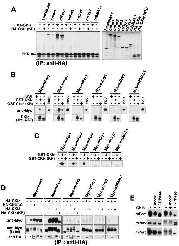
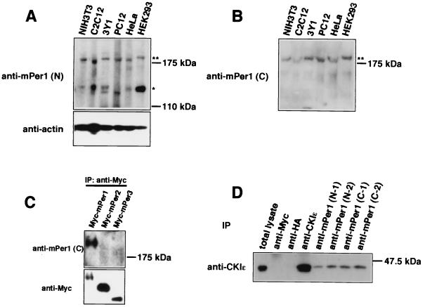
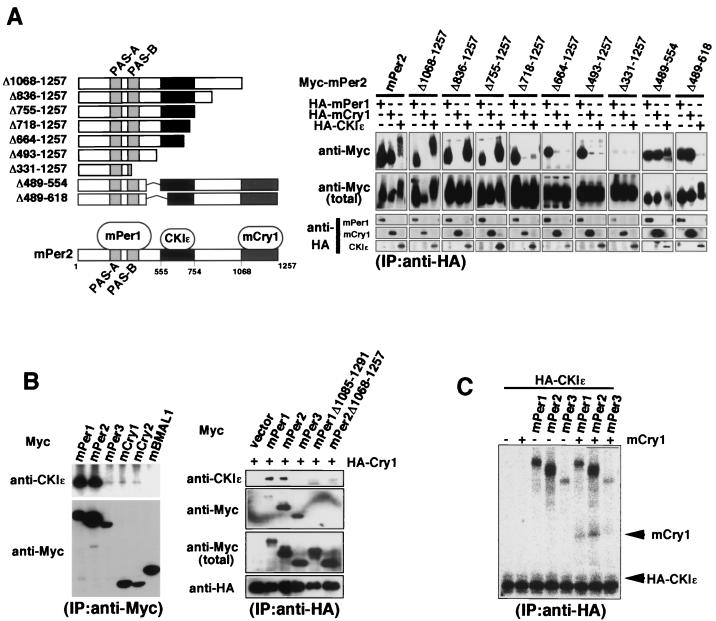
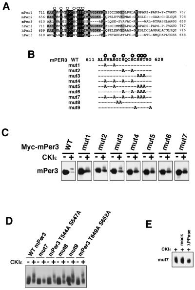
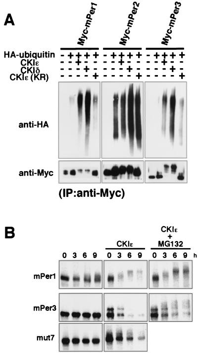
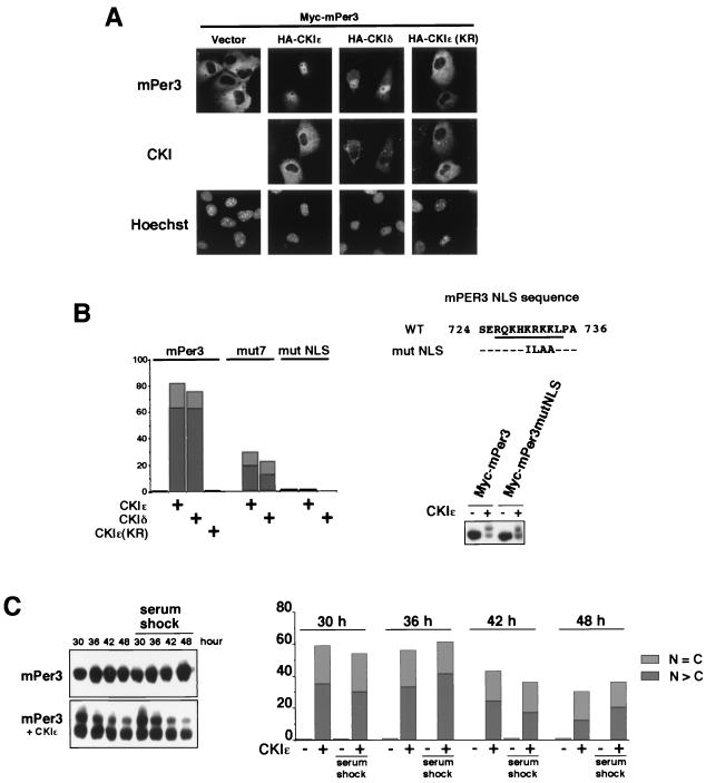
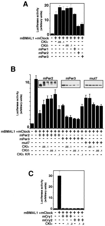
Recent studies have shown that casein kinase I epsilon (CKIepsilon) is an essential regulator of the mammalian circadian clock. However, the detailed mechanisms by which CKIepsilon regulates each component of the circadian negative-feedback loop have not been fully defined. We show here that mPer proteins, negative limbs of the autoregulatory loop, are specific substrates for CKIepsilon and CKIdelta. The CKI phosphorylation of mPer1 and mPer3 proteins results in their rapid degradation, which is dependent on the ubiquitin-proteasome pathway. Moreover, CKIepsilon and CKIdelta are able to induce nuclear translocation of mPer3, which requires its nuclear localization signal. The mutation in potential phosphorylation sites on mPer3 decreased the extent of both nuclear translocation and degradation of mPer3 that are stimulated by CKIepsilon. CKIepsilon and CKIdelta affected the inhibitory effect of mPer proteins on the transcriptional activity of BMAL1-CLOCK, but the inhibitory effect of mCry proteins on the activity of BMAL1-CLOCK was unaffected. These results suggest that CKIepsilon and CKIdelta regulate the mammalian circadian autoregulatory loop by controlling both protein turnover and subcellular localization of mPer proteins.


Figures

FIG. 1.
FIG. 1.
CKIɛ and CKIδ bind to and phosphorylate mPer proteins specifically. (A) Luciferase (control), mPer1, mPer2, mPer3, mCry1, mCry2, and mBMAL1 were separately synthesized in vitro by using TNT T7 Coupled Reticulocyte Lysate System (right panel) and then mixed with in vitro-synthesized HA-tagged CKIɛ or CKIɛ(KR) and incubated. Each of these mixtures was subjected to immunoprecipitation with anti-HA antibody (12CA5). After resolution by SDS-PAGE, immunoprecipitates were detected by a Bio-Rad phosphorimager (left panel). (B) Myc-tagged mPer1, mPer2, mPer3, mCry1, mCry2, or mBMAL1 was expressed in COS7 cells. Each of the cell extracts was incubated with GST, GST-fused CKIɛ, or GST-fused CKIɛ(KR) (expressed in E. coli, and purified) and glutathione-Sepharose 4B. The beads were then washed with the incubation buffer. After resolution by SDS-PAGE, proteins were analyzed by immunoblotting with anti-Myc antibody (A-14). (C) Myc-tagged mPer1, mPer2, mPer3, mCry1, mCry2, or mBMAL1 was expressed in COS7 cells. The cell extracts were subjected to immunoprecipitation with anti-Myc antibody (9E10). The washed beads were mixed with a kinase reaction buffer supplemented with GST-fused CKIɛ or GST-fused CKIɛ(KR), as well as [γ-32P]ATP, and incubated for 30 min. After resolution by SDS-PAGE, substrate phosphorylation was detected by a Bio-Rad phosphorimager. (D) Myc-tagged mPer1, mPer2, mPer3, mCry1, mCry2, or mBMAL1 was expressed in COS7 cells, together with HA-tagged CKIɛ, CKIɛΔC, CKIδ, or CKIɛ(KR). The cell lysates were subjected to immunoprecipitation with anti-HA antibody (12CA5). After resolution by SDS-PAGE, immunoprecipitates were analyzed by immunoblotting with anti-Myc antibody (A-14). (E) Myc-tagged mPer1, mPer2, or mPer3 was expressed in COS7 cells, together with or without CKIδ. The cell lysates were subjected to immunoprecipitation with anti-Myc antibody (9E10), and the washed beads were resuspended in phosphatase buffer and incubated with or without (mock) 40 U of purified lambda phosphatase for 30 min. After resolution by SDS-PAGE, the immunoprecipitates were analyzed by immunoblotting with anti-Myc antibody (A-14).
FIG. 2.
FIG. 2.
In vivo association between mPer1 and CKIɛ. (A and B) Various mammalian cells were lysed in the incubation buffer. After the cell extracts were resolved by SDS-PAGE, they were analyzed by immunoblotting with anti-mPer1(N) antibody or anti-actin antibody (loading control) (A) or with anti-mPer1(C) antibody (B). (C) Myc-tagged mPer1, mPer2, or mPer3 was expressed in COS7 cells, and the cell lysates were subjected to immunoprecipitation with anti-Myc antibody (9E10). These immunoprecipitates were analyzed by immunoblotting with anti-mPer1(C) antibody (upper panel) or anti-Myc antibody (A-14) (lower panel). (D) For coimmunoprecipitation experiments, NIH 3T3 cells were incubated in hypotonic lysis buffer and homogenized. Each of antibodies and protein G-Sepharose beads were added to the resulting supernatant. The beads were then washed with the hypotonic lysis buffer. The immunoprecipitates were analyzed by immunoblotting with anti-CKIɛ antibody. Anti-HA antibody and anti-Myc antibody were used as a nonrelevant antibody control.
FIG. 3.
FIG. 3.
mPer, mCry, and CKIɛ form a ternary complex. (A) In the left panel is a diagram of the deletion constructs of mPer2 and a summary of the results of the coimmunoprecipitation experiments (a CKIɛ binding domain and an mCry1 binding domain are indicated by dark gray and light gray boxes, respectively). In the right panel a CKIɛ binding domain and an mCry1 binding domain on mPer2 are identified. For coimmunoprecipitation experiments, Myc-tagged mPer2 or each of various fragments of mPer2 was expressed in COS7 cells, together with HA-tagged mPer1, mCry1, or CKIɛ. HA-tagged mPer1, mCry1, or CKIɛ was immunoprecipitated with anti-HA antibody (12CA5). The immunoprecipitates were analyzed by immunoblotting with anti-Myc antibody (A-14). (B, left panel) For the coimmunoprecipitation of endogenous CKIɛ, Myc-tagged mPer1, mPer2, mPer3, mCry1, mCry2, or mBMAL1 was expressed in COS7 cells. The cell lysates were subjected to immunoprecipitation with anti-Myc antibody (A-14). After resolution by SDS-PAGE, the immunoprecipitates were analyzed by immunoblotting with anti-CKIɛ antibody. (B, right panel) mPer, mCry, and CKIɛ form a ternary complex in cultured cells. Myc-tagged mPer1, mPer2, mPer3, mPer1Δ1085-1291, or mPer2Δ1068-1257 was expressed in COS7 cells, together with HA-tagged mCry1. The cell lysates were subjected to immunoprecipitation with anti-HA polyclonal antibody. After resolution by SDS-PAGE, the immunoprecipitates were analyzed by immunoblotting with anti-CKIɛ antibody. (C) mPer, mCry, and CKIɛ form a ternary complex in vitro. mPer1, mPer2, mPer3, mCry1, or HA-tagged CKIɛ was separately synthesized in vitro by using TNT T7 Coupled Reticulocyte Lysate System. mPer1, mPer2, or mPer3 was then mixed with HA-tagged CKIɛ, together with or without mCry1, and incubated. Each of these mixtures was then immunoprecipitated with anti-HA antibody (12CA5). The immunoprecipitates were detected with a Bio-Rad phosphorimager.
FIG. 4.
FIG. 4.
Identification of potential CKIɛ phosphorylation sites on mPer3. (A) Amino acid sequences of the mammalian Per family. Amino acid identities and similarities are indicated by dark gray and light gray boxes, respectively. The conserved serine-threonine residues are indicated by open circles. (B) PCR-generated alanine mutations were made in the mPer3 conserved serine-threonine cluster and compared with the wild-type (WT) mPer3 sequence. (C and D) Wild-type (WT) mPer3 or each of various mutants of mPer3 (0.7 μg of DNA) was expressed in COS7 cells, together with or without CKIɛ (0.3 μg of DNA). The cell extracts were subjected to immunoblotting with anti-Myc antibody (A-14). Equal amounts of proteins were loaded. The electrophoretic retardation of the mPer3 protein bands results from phosphorylation. (E) Myc-tagged mut7 was expressed in COS7 cells, together with or without CKIɛ. The cell lysates were subjected to immunoprecipitation with anti-Myc antibody (9E10) and incubated with or without (mock) purified lambda phosphatase. The immunoprecipitates were analyzed by immunoblotting with anti-Myc antibody (A-14).
FIG. 5.
FIG. 5.
CKI promotes ubiquitination of mPer proteins. (A) To show ubiquitination of mPer proteins, each of Myc-tagged mPer proteins was coexpressed with HA-tagged ubiquitin in the presence or absence of CKIɛ, CKIδ, or CKIɛ(KR). The cell lysates were then subjected to immunoprecipitation with anti-Myc antibody (9E10). After resolution by SDS-PAGE, immunoprecipitates were analyzed by immunoblotting with anti-HA polyclonal antibody. The accumulation of high-molecular-mass species recognized by the anti-HA antibody indicates that Myc-tagged proteins become multiply ubiquitinated. (B) To study the metabolic stability of mPer proteins, each of Myc-tagged mPer proteins was expressed in COS7 cells, together with or without CKIɛ. The cells were incubated in methionine-cysteine-deficient medium and then pulsed with 35S-labeled methionine-cysteine. The cells were incubated for the indicated lengths of time in DMEM-10% fetal calf serum with or without MG-132. The cell lysates were subjected to immunoprecipitation with anti-Myc antibody (9E10). The immunoprecipitates were detected with a Bio-Rad phosphorimager.
FIG. 6.
FIG. 6.
CKI-induced nuclear translocation of mPer3 depends on CKI-mediated phosphorylation and NLS of mPer3. (A) Myc-tagged mPer3 was expressed in COS7 cells, together with or without HA-tagged CKIɛ, CKIδ or CKIɛ(KR). The cells were fixed and incubated with primary antibodies (anti-Myc antibody [9E10] and anti-HA polyclonal antibody) and then with the appropriate secondary antibodies. The cells were examined by using a Zeiss Axiophoto. (B, left panel) Myc-tagged mPer3, mut7, or mutNLS (see the right panel) (0.9 μg of DNA) was expressed in COS7 cells, together with or without HA-tagged CKIɛ, CKIδ, or CKIɛ(KR) (0.1 μg of DNA). After being stained, the cells were classified into three categories in terms of location of Myc-tagged mPer proteins as nucleus (N > C), cytoplasm (N < C), or both nucleus and cytoplasm (N = C). The classifications “N > C” and “N = C” are indicated by dark gray and light gray boxes, respectively. More than 150 cells were examined, and the percentages of N > C and N = C are shown. Experiments were performed twice with similar results. (B, right panel) A PCR-generated mutation was made in a presumable mPer3 NLS (mutNLS) and compared to the wild-type (WT) sequence. WT mPer3 or mPer3mutNLS was expressed in COS7 cells, together with or without CKIɛ. The cell extracts were subjected to immunoblotting with anti-Myc antibody (A-14). (C) The cells were subjected to transfection and kept in a medium containing 1% serum, after the serum shock (50% for 2 h). Myc-tagged mPer3 was expressed in COS7 cells, together with or without CKIɛ. The cells were then lysed (or fixed for staining) 30, 36, 42, and 48 h after the serum shock (time = 0 h). In the left panel, cell extracts were subjected to immunoblotting with anti-Myc antibody (A-14). The electrophoretic retardation of the mPer3 protein bands results from phosphorylation. In the right panel, cells were classified after being stained into three categories with regard to the location of Myc-tagged mPer3: N > C, N < C, or N = C. The N > C and N = C classifications are indicated by dark gray and light gray boxes, respectively. More than 250 cells were examined, and the percentages of the N > C and N = C groups are shown.
FIG. 7.
FIG. 7.
The effect of CKI on the E-box-mediated transcription is seen only in the presence of mPer proteins, and the inhibitory effects of mPer2 and mPer3 are differently affected by CKI. The amounts of the constructs transfected varied depending on the experiment: mBMAL1 (100 ng), mClock (100 ng), and mPer (700 ng) (A and B) and mBMAL1 (250 ng), mClock (250 ng), and mCry (400 ng) (C). The amount of CKI expression vector (in micrograms) is shown in each panel. The total amount of DNA per well was adjusted to 1.1 μg by adding pcDNA3 vector as the carrier. At 36 h after transfection, COS7 cells were scraped into 150 μl of reporter lysis buffer and then centrifuged. The supernatant was used to detect the luciferase activity. The data were normalized for β-galactosidase activity. The relative luciferase activities (means ± the standard deviations; n = 3) are presented. These experiments were repeated three times, and the data shown are representative. (A) The effect of CKIɛ, CKIδ, mPer1, mPer2, or mPer3 on mBMAL1-mCLOCK-mediated transcription. (B) CKIɛ and CKIδ reduce the inhibitory effect of mPer2 on the transcriptional activity of BMAL1-CLOCK but enhance the inhibitory effect of mPer3. The amounts of the mPer proteins are shown (upper panel). mut7 is one of the potential phosphorylation site mutants of mPer3 (see Fig. 4B). The data for the effects of CKIɛ(KR) on the inhibitory effect of mPer proteins were normalized, since CKIɛ(KR) suppressed the transcriptional activity of BMAL1-CLOCK to some extent (data not shown). (C) CKIɛ or CKIδ does not affect the inhibitory effect of mCry1 and mCry2 on the transcriptional activity of BMAL1-CLOCK.

References

    1. Albrecht, U., Z. S. Sun, G. Eichele, and C. C. Lee. 1997. A differential response of two putative mammalian circadian regulators, mPer1 and mPer2, to light. Cell 91:1055-1064. - PubMed
    1. Antoch, M. P., E. J. Song, A. M. Chang, M. H. Vitaterna, Y. Zhao, L. D. Wilsbacher, A. M. Sangoram, D. P. King, L. H. Pinto, and J. S. Takahashi. 1997. Functional identification of the mouse circadian Clock gene by transgenic BAC rescue. Cell 89:655-667. - PMC - PubMed
    1. Brown, S. A., and U. Schibler. 1999. The ins and outs of circadian timekeeping. Curr. Opin. Genet. Dev. 9:588-594. - PubMed
    1. Darlington, T. K., K. Wager-Smith, M. F. Ceriani, D. Staknis, N. Gekakis, T. D. Steeves, C. J. Weitz, J. S. Takahashi, and S. A. Kay. 1998. Closing the circadian loop: CLOCK-induced transcription of its own inhibitors per and tim. Science 280:1599-1603. - PubMed
    1. Devlin, P. F., and S. A. Kay. 1999. Cryptochromes—bringing the blues to circadian rhythms. Trends Cell Biol. 9:295-298. - PubMed

Publication types

MeSH terms

LinkOut - more resources