. 2002 Mar;22(6):1704-13.
doi: 10.1128/MCB.22.6.1704-1713.2002.

The endothelial receptor tyrosine kinase Tie1 activates phosphatidylinositol 3-kinase and Akt to inhibit apoptosis

Affiliations

The endothelial receptor tyrosine kinase Tie1 activates phosphatidylinositol 3-kinase and Akt to inhibit apoptosis

Christopher D Kontos et al. Mol Cell Biol. 2002 Mar.

Abstract

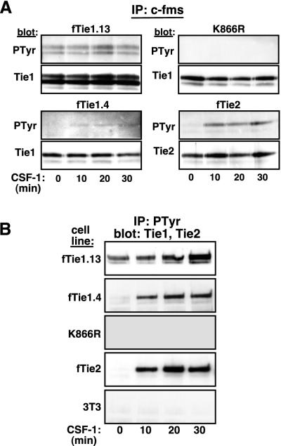
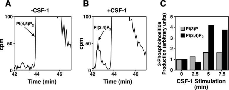
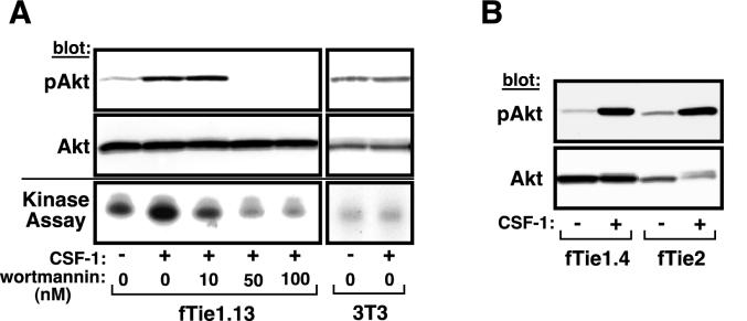
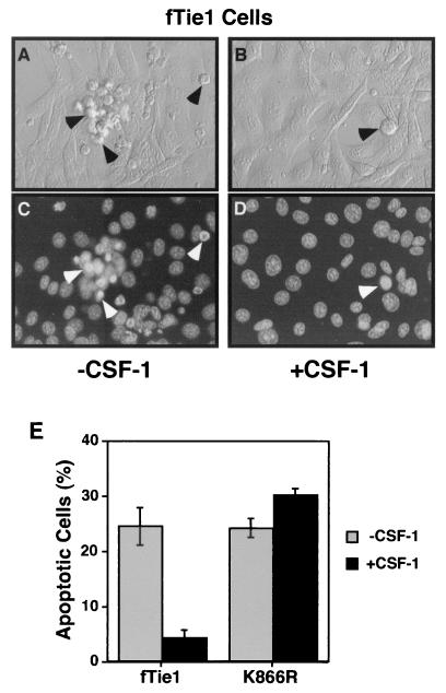
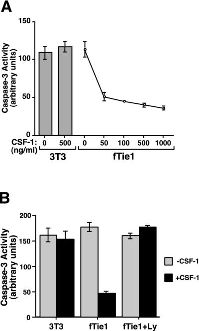
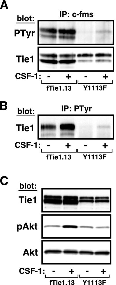
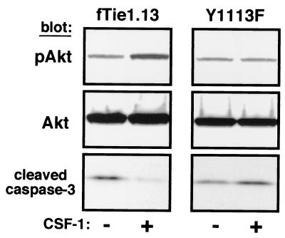
Tie1 is an orphan receptor tyrosine kinase that is expressed almost exclusively in endothelial cells and that is required for normal embryonic vascular development. Genetic studies suggest that Tie1 promotes endothelial cell survival, but other studies have suggested that the Tie1 kinase has little to no activity, and Tie1-mediated signaling pathways are unknown. To begin to study Tie1 signaling, a recombinant glutathione S-transferase (GST)-Tie1 kinase fusion protein was produced in insect cells and found to be autophosphorylated in vitro. GST-Tie1 but not a kinase-inactive mutant associated with a recombinant p85 SH2 domain protein in vitro, suggesting that Tie1 might signal through phosphatidylinositol (PI) 3-kinase. To study Tie1 signaling in a cellular context, a c-fms-Tie1 chimeric receptor (fTie1) was expressed in NIH 3T3 cells. Ligand stimulation of fTie1 resulted in Tie1 autophosphorylation and downstream activation of PI 3-kinase and Akt. Stimulation of fTie1-expressing cells potently inhibited UV irradiation-induced apoptosis in a PI 3-kinase-dependent manner. Moreover, both Akt phosphorylation and inhibition of apoptosis were abrogated by mutation of tyrosine 1113 to phenylalanine, suggesting that this residue is an important PI 3-kinase binding site. These findings are the first biochemical demonstration of a signal transduction pathway and corresponding cellular function for Tie1, and the antiapoptotic effect of Tie1 is consistent with the results of previous genetic studies.


Figures

FIG. 1.
FIG. 1.
p85 associates with Tie1 in vitro. (A) Comparison of Tie1 and Tie2 C-terminal amino acid sequences. C-terminal regions of Tie1 and Tie2 containing putative autophosphorylation sites were aligned and compared to the consensus p85-binding motif. The greatest difference in this region is in the Y + 3 position of the putative p85-binding motif. Key tyrosine residues and the putative p85-binding motif within each kinase are in boldface. Positions of tyrosine residues within each kinase are shown above and below the alignment. Solid lines, identical amino acid residues; dashed lines, conserved residues. Residues are abbreviated according to the single-letter amino acid code. (B) Tie1 is autophosphorylated and associates with p85 in vitro. Wild-type GST-Tie1 and a kinase-inactive mutant (K866R) were purified on glutathione Sepharose from Sf9 insect cell lysates infected with recombinant baculoviruses encoding each protein. As a control, an equal volume of uninfected Sf9 cell lysate was incubated with glutathione Sepharose (beads). Following an in vitro kinase reaction, the samples were incubated with recombinant C-terminal p85 SH2 domain protein. The GST kinases and associated proteins were eluted and analyzed by SDS-8 to 16% PAGE and Western blotting with anti-Tie1, antiphosphotyrosine (PTyr), and anti-p85 Abs. Similar results were obtained in three separate experiments. Note that equal starting amounts of GST-Tie1-wild type and -K866R were confirmed by Coomassie staining of glutathione Sepharose-purified Sf9 lysates.
FIG. 2.
FIG. 2.
Tie1 is autophosphorylated in vivo. (A) Autophosphorylation of Tie1 is kinase dependent. Two independent clones of NIH 3T3 cells expressing wild-type fTie1 as well as cells expressing a kinase-inactive mutant of fTie1 (K866R) or fTie2 were serum starved and were then treated with CSF-1 (500 ng/ml) for 0, 10, 20, or 30 min. Lysates from these cells were immunoprecipitated with anti-c-fms, separated by SDS-8% PAGE, and Western blotted with antiphosphotyrosine (PTyr, clone PY99), and then stripped and reprobed with anti-Tie1 or anti-Tie2. Weak but detectable increases in ligand-induced tyrosine phosphorylation were observed in both fTie1-expressing clones but not in K866R cells. As a control, fTie2 cells showed readily detectable ligand-induced autophosphorylation. (B) Tie1 is present in antiphosphotyrosine immunoprecipitates. The cells used for panel A, as well as untransfected 3T3 cells, were treated as described above, except lysates were immunoprecipitated with antiphosphotyrosine (PY99) and were then Western blotted with anti-Tie1 or (for fTie2 cell lysates) anti-Tie2. Tie1 was readily detectable in antiphosphotyrosine immunoprecipitates from both low- and high-receptor-expressing fTie1 cells but not from K866R cells or untransfected 3T3 cells. CSF-1 treatment also resulted in the presence of Tie2 in phosphotyrosine immunoprecipitates.
FIG. 3.
FIG. 3.
Tie1 activates PI 3-kinase in vivo. (A and B) NIH 3T3 cells expressing fTie1 were labeled 72 h with 12.5 μCi of [3H]myo-inositol/ml and were then serum starved for 3 h. Cells were left untreated (A) or were treated with CSF-1 (500 ng/ml) for 2.5, 5, or 7.5 min (B), all in the presence of 1 mM vanadate, and lipids were extracted and separated by HPLC on a strong anion-exchange column. A peak corresponding to PI 3,4-bisphosphate [PI(3,4)P2] was detected at 5 and 7.5 min (shown) after CSF-1 treatment. The regions of the chromatograms where PI(3,4)P2 and PI(4,5)P2 eluted (between 42 and 47 min) are shown, and the peaks corresponding to PI(4,5)P2, which is much more abundant than PI(3,4)P2, are truncated at 100 cpm (maximum values were approximately 40,000 to 60,000 cpm). (C) Quantification of 3-phosphoinositide production by Tie1. On each chromatogram, total counts per minute under the peaks corresponding to PI(3)P (not shown) and PI(3,4)P2 were quantified and expressed relative to the total PI from that sample. CSF-1 induced a maximal 1.6-fold increase in PI(3)P and a 4.2-fold increase in PI(3,4)P2. Similar results were obtained in a duplicate experiment.
FIG. 4.
FIG. 4.
Tie1 induces phosphorylation and activation of Akt. (A) fTie1-expressing cells or untransfected NIH 3T3 cells were serum starved overnight and were then left untreated or were treated 8 min with CSF-1 (500 ng/ml) in the presence of 1 mM vanadate and the indicated concentration of wortmannin or an equal volume of DMSO. An aliquot of each cell lysate was separated by SDS-8% PAGE and Western blotted with Abs specific for phosphoserine 473 of Akt (pAkt) or total Akt (upper panels). The remainder of each sample was immunoprecipitated with anti-Akt and was used in an in vitro kinase reaction with [γ-32P]ATP and recombinant histone H2B as a substrate. Radiolabeled histone H2B was separated by SDS-15% PAGE and evaluated by autoradiography (lower panel). (B) A second independent clone of fTie1-expressing cells as well as fTie2 and K866R cells (not shown) was treated as described for panel A, and cell lysates were Western blotted sequentially with anti-phospho-Akt and anti-Akt.
FIG. 5.
FIG. 5.
Tie1 blocks UV irradiation-induced apoptosis. NIH 3T3 cells expressing fTie1.13 (wild-type) or the kinase-inactive mutant (K866R) were grown 24 h in 60-mm-diameter dishes and were then serum starved 15 h in the absence or presence of CSF-1 (500 ng/ml). The cells were then UV irradiated with 1,200 J/m2, stained with Hoechst 33342 (10 μg/ml) and propidium iodide (2.5 μg/ml), and evaluated by phase-contrast (A and B) and fluorescence microscopy (C and D). Untreated fTie1 cells (A and C) began to display morphological characteristics of apoptosis, including membrane blebbing and nuclear condensation (arrowheads), as early as 1 h after UV irradiation, whereas CSF-1-treated fTie1 cells (B and D) remained viable. (E) Quantification of apoptosis following UV irradiation. The number of apoptotic nuclei was counted after Hoechst staining and expressed as the percentage of total nuclei ± standard error of the mean. CSF-1 treatment significantly decreased apoptosis of wild-type cells but not of K866R mutant cells.
FIG. 6.
FIG. 6.
Tie1 inhibits UV irradiation-induced caspase 3 activation through a PI 3-kinase-dependent mechanism. (A) Untransfected NIH 3T3 cells or 3T3 cells expressing fTie1 were grown in 60-mm-diameter dishes for 24 h and then serum starved 15 h in the absence or presence of the indicated concentration of CSF-1 and were then UV irradiated as described for Fig. 5. Three hours after UV irradiation, cells were lysed and the lysates were used in a colorimetric assay of caspase 3 activity (ApoAlert; Clontech). fTie1 blocked the UV-induced increase in caspase 3 activity in a dose-dependent manner. (B) Untransfected 3T3 cells and fTie1 cells were serum starved 15 h with or without 500 ng of CSF-1/ml and Ly294002 (Ly) (30 μM) or an equal volume of DMSO. Caspase 3 activity was analyzed as for panel A. The inhibition of caspase 3 activity by Tie1 was completely reversed by Ly294002.
FIG. 7.
FIG. 7.
Mutation of tyrosine 1113 abrogates Akt phosphorylation. (A and B) fTie1-Y1113F is functionally active. NIH 3T3 cells expressing either wild-type fTie1 (fTie1.13) or fTie1-Y1113F (Y1113F) were serum starved overnight and were then either left untreated or treated with CSF-1 (500 ng/ml) for 20 min. Receptors were immunoprecipitated from cell lysates with anti-c-fms (A) or antiphosphotyrosine (B). Proteins for panel A were Western blotted sequentially with antiphosphotyrosine and anti-Tie1, and proteins for panel B were Western blotted with anti-Tie1. Tyrosine phosphorylation of fTie1-Y1113F and its presence in phosphotyrosine immunoprecipitates were detected only after CSF-1 treatment. (C) The fTie1-Y1113F mutant fails to activate Akt. Cells expressing wild-type fTie1 or Y1113F were serum starved overnight and were then treated with or without CSF-1, all in the presence of vanadate (1 mM). An aliquot of each cell lysate was separated by SDS-8% PAGE and Western blotted with the indicated Abs. CSF-1 stimulation of Y1113F cells failed to induce Akt phosphorylation.
FIG. 8.
FIG. 8.
Mutation of Y1113 blocks the antiapoptotic effect of Tie1. Cells expressing wild-type fTie1 or Y1113F were serum starved overnight in the absence or presence of CSF-1 (500 ng/ml). The following morning the cells were UV irradiated (1,200 J/m2). Cell lysates were separated by SDS-8 to 16% PAGE and Western blotted with the indicated Abs. CSF-1 treatment induced an increase in Akt phosphorylation and a decrease in caspase 3 cleavage in wild-type fTie1 cells but not in Y1113F-expressing cells.

References

    1. Carpenter, C. L., and L. C. Cantley. 1996. Phosphoinositide kinases. Curr. Opin. Cell Biol. 8:153-158. - PubMed
    1. Davis, S., T. H. Aldrich, P. F. Jones, A. Acheson, D. L. Compton, V. Jain, T. E. Ryan, J. Bruno, C. Radziejewski, P. C. Maisonpierre, and G. D. Yancopoulos. 1996. Isolation of angiopoietin-1, a ligand for the Tie-2 receptor, by secretion-trap expression cloning. Cell 87:1161-1169. - PubMed
    1. Downing, J. R., M. F. Roussel, and C. J. Sherr. 1989. Ligand and protein kinase C downmodulate the colony-stimulating factor 1 receptor by independent mechanisms. Mol. Cell. Biol. 9:2890-2896. - PMC - PubMed
    1. Downward, J. 1998. Mechanisms and consequences of activation of protein kinase B/Akt. Curr. Opin. Cell Biol. 10:262-267. - PubMed
    1. Dubois, N. A., L. C. Kolpack, R. Wang, R. G. Azizkhan, and V. L. Bautch. 1991. Isolation and characterization of an established endothelial cell line from transgenic mouse hemangiomas. Exp. Cell Res. 196:302-313. - PubMed

Publication types

MeSH terms

Substances