. 2002 Mar;22(6):1754-66.
doi: 10.1128/MCB.22.6.1754-1766.2002.

IAP suppression of apoptosis involves distinct mechanisms: the TAK1/JNK1 signaling cascade and caspase inhibition

Affiliations

IAP suppression of apoptosis involves distinct mechanisms: the TAK1/JNK1 signaling cascade and caspase inhibition

M Germana Sanna et al. Mol Cell Biol. 2002 Mar.

Abstract

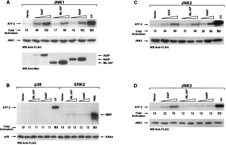
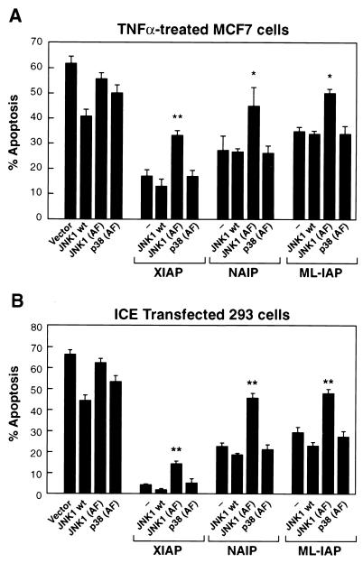
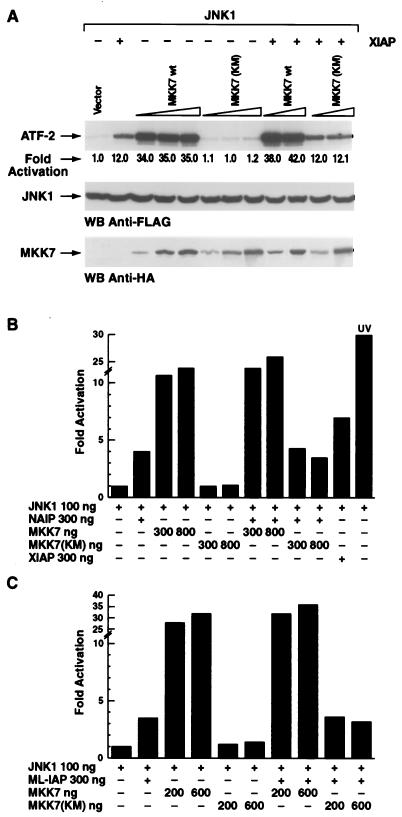
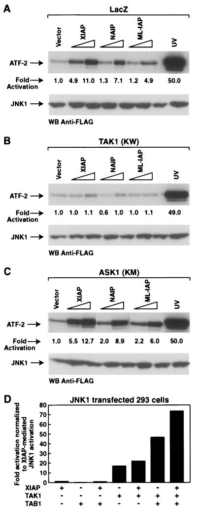
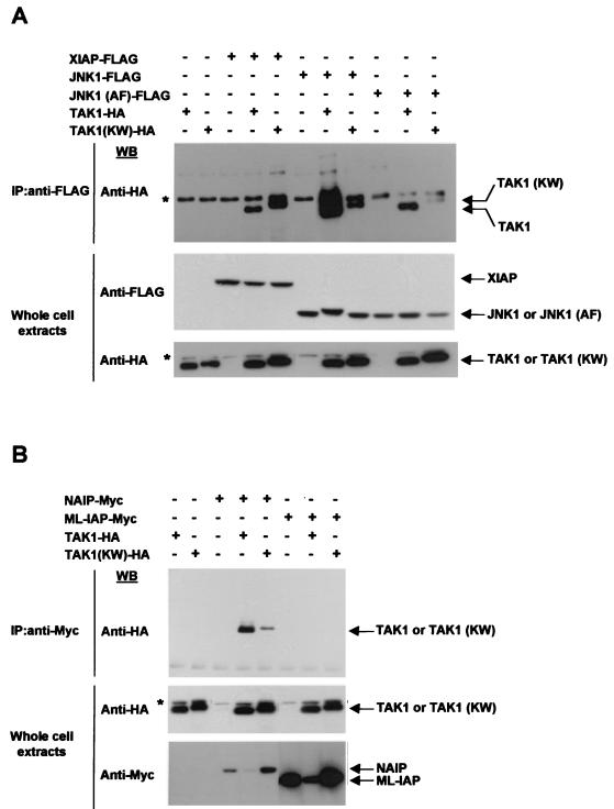
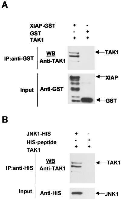
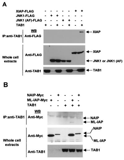
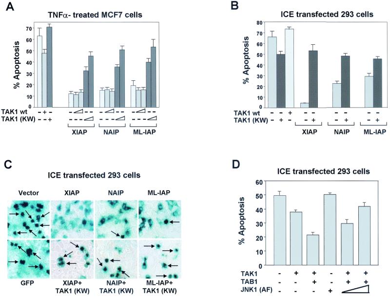
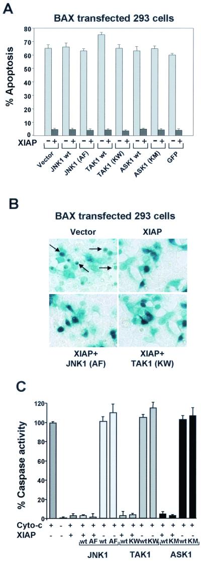
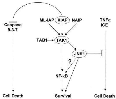
The antiapoptotic properties of the inhibitor of apoptosis (IAP) family of proteins have been linked to caspase inhibition. We have previously described an alternative mechanism of XIAP inhibition of apoptosis that depends on the selective activation of JNK1. Here we report that two other members of the IAP family, NAIP and ML-IAP, both activate JNK1. Expression of catalytically inactive JNK1 blocks NAIP and ML-IAP protection against ICE- and TNF-alpha-induced apoptosis, indicating that JNK1 activation is necessary for the antiapoptotic effect of these proteins. The MAP3 kinase, TAK1, appears to be an essential component of this antiapoptotic pathway since IAP-mediated activation of JNK1, as well as protection against TNF-alpha- and ICE-induced apoptosis, is inhibited when catalytically inactive TAK1 is expressed. In addition, XIAP, NAIP, and JNK1 bind to TAK1. Importantly, expression of catalytically inactive TAK1 did not affect XIAP inhibition of caspase activity. These data suggest that XIAP's antiapoptotic activity is achieved by two separate mechanisms: one requiring TAK1-dependent JNK1 activation and the second involving caspase inhibition.


Figures

FIG. 1.
FIG. 1.
XIAP, NAIP, and ML-IAP selectively activate JNK kinases. 293T cells were transfected with vectors encoding JNK1 (A), p38 or ERK2 (B), JNK2 (C), or JNK3 (D) (200 ng each) in the absence or presence of increasing concentrations of XIAP, NAIP-BIR1-3, or ML-IAP (200 or 800 ng). The amount of transfected cDNA was kept constant in each sample by adding control pcDNA3 vector. An in vitro kinase assay was performed using ATF-2 or MBP as substrate. Kinase activity was quantitated by PhosphorImager and is expressed as fold induction relative to the basal level of phosphorylation of each MAP kinase. UV and phorbol myristate acetate were used as positive controls. Western blottings showing equal expression levels of JNK1 are reported for each experiment. Expression levels of XIAP, NAIP-BIR1-3, or ML-IAP were comparable in each experiment and therefore are shown only in panel A.
FIG. 2.
FIG. 2.
XIAP, NAIP, and ML-IAP require JNK1 for protection against apoptosis. (A) Effect of wt JNK1, JNK1 (AF), or p38 (AF) on XIAP, NAIP, or ML-IAP protection against TNF-α-induced apoptosis. MCF7 cells were transfected with control vector pcDNA3 alone or plasmids encoding XIAP, NAIP-BIR1-3, or ML-IAP (200 ng each), together with control vector, wt JNK1, JNK1 (AF), or p38 (AF) as a control (800 ng each). Transfected cells were treated with TNF-α for 12 h (100 ng/ml) and processed as described in Materials and Methods. Effects on cell viability were determined using X-Gal staining and AnnexinV-PE/FACS analysis. % apoptosis, incidence of apoptotic cells among the β-Gal-positive cells (transfected cells). Data represent the mean ± standard error of at least three experiments, each run in duplicate and scored blind. ∗, P < 0.05 (determined by an unpaired t test); ∗∗, P < 0.001. Values were calculated compared to the control samples expressing only pcDNA3. (B) Effect of wt JNK1 or JNK1 (AF) on XIAP, NAIP, or ML-IAP protection against ICE-induced apoptosis. 293T cells were transfected with plasmids encoding ICE-β-Gal alone (200 ng) or together with wt JNK1, JNK1 (AF), or p38 (AF) alone or in the presence of pcDNA3, XIAP, NAIP-BIR1-3, or ML-IAP (200 ng each). Parental pcDNA3 vector was added to normalize the amount of transfected DNA.
FIG. 3.
FIG. 3.
XIAP, NAIP, and ML-IAP activate JNK1 independently of MKK7/MKK4. Increasing amounts (from 200 to 800 ng) of wt MKK7 or MKK7 (KM), were cotransfected together with JNK1 (100 ng) in the presence or absence of XIAP (A), NAIP (B), or ML-IAP (C) (200 ng each). In vitro kinase assays were performed on immunoprecipitated JNK1 using ATF-2 as substrate, and kinase activity was quantitated by PhosphorImager. Western blottings show consistent expression of JNK1 and wt MKK7 or MKK7 (KM) using anti-FLAG and anti-HA antibody, respectively. UV activation is shown as a positive control of JNK1 activation. The standard deviation was always <6%.
FIG. 4.
FIG. 4.
XIAP, NAIP, and ML-IAP activate JNK1 through TAK1. (A, B, C) Effects of LacZ, TAK1 (KW), or ASK1 (KM) on XIAP-, NAIP-, and ML-IAP-dependent JNK1 activation. Plasmids encoding wild-type JNK1 (100 ng) and increasing amounts of XIAP, NAIP-BIR1-3, or ML-IAP (200 and 600 ng) were transfected in 293T cells stably expressing LacZ control gene (A), TAK1 (KW) (B), or ASK1 (KM) (C). An in vitro kinase assay was performed on immunoprecipitated JNK1 using ATF-2 as substrate, and kinase activity was quantitated by PhosphorImager. UV stimulation is also shown. Western blottings show equal expression of JNK1. Expression of XIAP, NAIP, or ML-IAP was not altered by the expression of TAK1 (KW) (data not shown). (D) Effects of XIAP, TAK1, and TAB1 on JNK1 activation. 293T cells were transfected with vectors encoding JNK1 (100 ng) plus XIAP (600 ng), TAK1 (15 ng), or TAB1 (5 ng) alone or in combination. In vitro kinase assay was performed using ATF-2 as a substrate. The levels of kinase activity are expressed as fold induction normalized to the XIAP-mediated activation of JNK1.
FIG. 5.
FIG. 5.
XIAP, NAIP, and JNK1 interact with TAK1 in vivo. (A) In vivo interaction of XIAP, JNK1, or JNK1 (AF) with TAK1 or TAK1 (KW). Vectors encoding XIAP-FLAG, JNK1-FLAG, or JNK1 (AF)-FLAG were cotransfected with wt TAK1-HA or TAK1 (KW)-HA in 293T cells. Cell extracts were immunoprecipitated (IP) using anti-FLAG antibody-conjugated beads. Coprecipitated TAK1 or TAK1 (KW) was detected by Western blot analysis with an anti-HA antibody. Cell extracts were also directly subjected to immunoblot analysis (IB) to check for protein expression. Asterisks indicate the presence of an unspecific band that appears when the anti-HA antibody is used. (B) Interaction of NAIP or ML-IAP with TAK1 or TAK1 (KW). Vectors encoding NAIP-BIR1-3-Myc or ML-IAP-Myc were cotransfected with TAK1-HA or TAK1 (KW)-HA in 293T cells. Cell extracts were immunoprecipitated using anti-Myc antibody. Coprecipitated TAK1 or TAK1 (KW) was detected by Western blot analysis with an anti-HA antibody. Cells extracts were also directly subjected to immunoblot analysis to check for protein expression.
FIG. 6.
FIG. 6.
XIAP, NAIP, and JNK1 directly interact with TAK1. (A and B) In vitro interaction of XIAP or JNK1 with TAK1. GST-XIAP or HIS-JNK1 or the respective negative controls GST and HIS-peptide recombinant proteins were incubated with gluthatione or Ni-NTA-conjugated beads and in vitro-translated TAK1 protein was added. Coprecipitation of TAK1 was detected by Western blotting using an anti-TAK1 antibody. Input proteins were detected by Western blot using anti-GST and anti-HIS antibodies.
FIG. 7.
FIG. 7.
XIAP, NAIP, and ML-IAP interact with TAB1. (A) Interaction of XIAP, JNK1, or JNK1 (AF) with TAB1. Vectors encoding XIAP-FLAG, JNK1-FLAG, or JNK1 (AF)-FLAG were cotransfected with TAB1 in 293T cells. Cell extracts were subjected to immunoprecipitation with anti-TAB1 antibody, and coprecipitated XIAP, JNK1, or JNK1 (AF) was detected by Western blot analysis using anti-FLAG antibody. Cells extracts were subjected to immunoblot analysis to check protein expression. (B) Interaction of NAIP or ML-IAP with TAB1. Vectors encoding NAIP-BIR1-3-Myc or ML-IAP-Myc were cotransfected with TAB1 in 293T cells. Cell extracts were subjected to immunoprecipitation with anti-TAB1 antibody and precipitated NAIP or ML-IAP were detected by Western blot analysis with anti-Myc antibody. Cells extracts were subjected to immunoblot analysis to check protein expression.
FIG. 8.
FIG. 8.
TAK1 (KW) inhibits XIAP, NAIP, and ML-IAP protection against TNF-α- and ICE-induced apoptosis. (A) Effect of TAK1 or TAK1 (KW) on XIAP, NAIP, or ML-IAP protection against TNF-α-induced apoptosis. MCF7-Fas cells were transfected with plasmids encoding XIAP, NAIP-BIR1-3, or ML-IAP (200 ng each), together with control vector or increasing concentrations of wt TAK1 or TAK1 (KM) (200 and 800 ng). Plasmid expressing β-Gal (200 ng) was also transfected to allow quantitation of apoptotic cells. The effect of expression of empty vector, TAK1, or TAK1 (KW) (800 ng each) without IAPs is also shown. Cells were treated with TNF-α (100 ng/ml) and processed as described in Materials and Methods. Effects on cell viability were determined by X-Gal staining (shown) and AnnexinV-PE/FACS analysis (not shown) and produced similar results. % apoptosis, incidence of apoptotic cells among the β-Gal positive (transfected cells). Data represent the mean ± standard error of at least three experiments, each run in duplicate and scored blind. (B and C) Effect of TAK1 (KW) on XIAP, NAIP, or ML-IAP protection against ICE-induced apoptosis. 293T cells were transfected with plasmids encoding ICE-β-Gal (200 ng) in the absence or presence of wt TAK1, TAK1 (KW), XIAP, NAIP-BIR1-3, or ML-IAP (300 ng each). Parental pcDNA3 vector was added to normalize the amount of transfected DNA. Cell viability was determined by X-Gal staining and AnnexinV-PE/FACS analysis. Arrows indicate examples of apoptotic cells. (D) Effect of activated TAK1 on apoptosis. 293T cells were transfected with plasmids encoding ICE-β-Gal (100 ng) in the presence or absence wt TAK1 or TAB1 alone (50 ng each) or in combination with increasing concentrations of JNK1 (AF) (200 and 800 ng). Cell viability was determined by X-Gal staining and AnnexinV-PE/FACS analysis and produced similar results.
FIG. 9.
FIG. 9.
JNK1 (AF) and TAK1 (KW) do not inhibit XIAP, NAIP, and ML-IAP protection against BAX- or caspase-induced apoptosis. (A and B) Effect of JNK1 (AF), TAK1 (KW), or ASK1 (KM) on XIAP protection against BAX-induced apoptosis. 293T cells were transfected with plasmids encoding BAX (100 ng) or XIAP together with one of the following: wt JNK1, JNK1 (AF), TAK1, TAK1 (KW), ASK1, or ASK1 (KM) (600 ng each). Plasmid encoding GFP was also transfected with BAX alone or BAX plus XIAP as a control. Expression vector encoding β-Gal (50 ng) was transfected together with the different plasmids to allow morphological observation of the cells. Effects on cell viability were determined as described above. Arrows indicate examples of apoptotic cells. (C) Effect of JNK1 (AF), TAK1 (KW), or ASK1 (KM) on XIAP inhibition of caspase activity. 293T cells were seeded in 100-mm-diameter dishes and transfected with plasmids encoding XIAP alone (3 μg) or with one of the following: wt JNK1, TAK1, ASK1, JNK1 (AF), TAK1 (KW), or ASK1 (KM) (3 μg). Empty vector was also transfected alone as a control (6 μg). Cell extracts were prepared and cytochrome c was added to induce proteolytic processing of pro-caspase 3. Caspase activity was measured by monitoring the release of AFC DEVD-containing synthetic peptides.
FIG. 10.
FIG. 10.
Model for the activation of JNK1 by IAPs. IAP-mediated activation of JNK1 passes through TAK1 and promotes cell survival by inhibiting TNF-α- and ICE-induced apoptosis. This event is separate from XIAP inhibition of caspases (see text for details).

References

    1. Ambrosini, G., C. Adida, and D. C. Altieri. 1997. A novel anti-apoptosis gene, survivin, expressed in cancer and lymphoma. Nat. Med. 3:917-921. - PubMed
    1. Bernardi, P., L. Scorrano, R. Colonna, V. Petronilli, and F. Di Lisa. 1999. Mitochondria and cell death. Mechanistic aspects and methodological issues. Eur. J. Biochem. 264:687-701. - PubMed
    1. Chen, Y. R., and T. H. Tan. 2000. The c-Jun N-terminal kinase pathway and apoptotic signaling. Int. J. Oncol. 16:651-662. - PubMed
    1. Clem, R. J., and L. K. Miller. 1994. Control of programmed cell death by the baculovirus genes p35 and iap. Mol. Cell. Biol. 14:5212-5222. - PMC - PubMed
    1. Decaudin, D., I. Marzo, C. Brenner, and G. Kroemer. 1998. Mitochondria in chemotherapy-induced apoptosis: a prospective novel target of cancer therapy. Int. J. Oncol. 12:141-152. - PubMed

Publication types

MeSH terms

Substances