. 2002 Mar;22(6):1778-91.
doi: 10.1128/MCB.22.6.1778-1791.2002.

A novel and conserved protein-protein interaction domain of mammalian Lin-2/CASK binds and recruits SAP97 to the lateral surface of epithelia

Affiliations

A novel and conserved protein-protein interaction domain of mammalian Lin-2/CASK binds and recruits SAP97 to the lateral surface of epithelia

Seonok Lee et al. Mol Cell Biol. 2002 Mar.

Abstract

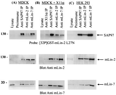
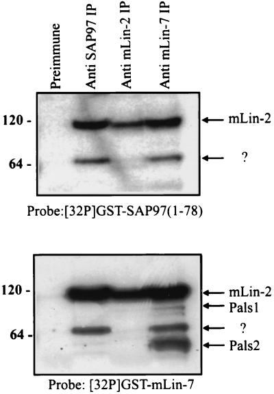
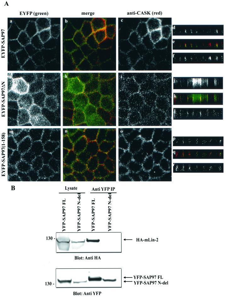
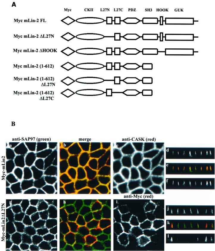
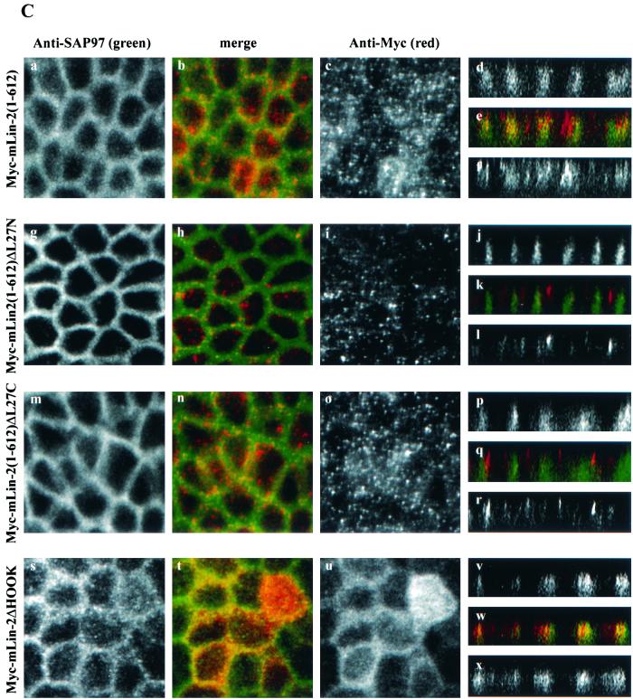
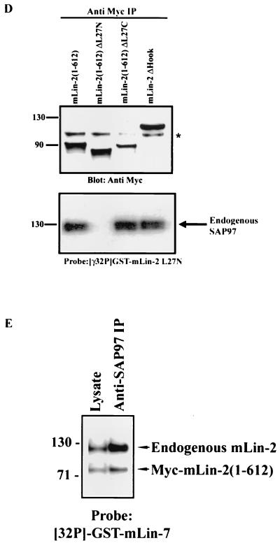
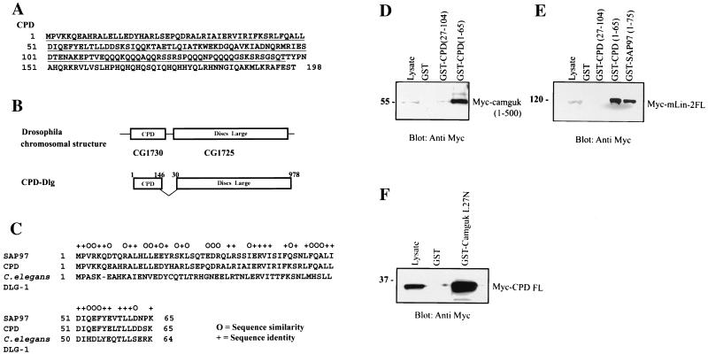
Mammalian Lin-2 (mLin-2)/CASK is a membrane-associated guanylate kinase (MAGUK) and contains multidomain modules that mediate protein-protein interactions important for the establishment and maintenance of neuronal and epithelial cell polarization. The importance of mLin-2/CASK in mammalian development is demonstrated by the fact that mutations in mLin-2/CASK or SAP97, another MAGUK protein, lead to cleft palate in mice. We recently identified a new protein-protein interaction domain, called the L27 domain, which is present twice in mLin-2/CASK. In this report, we further define the binding of the L27C domain of mLin-2/CASK to the L27 domain of mLin-7 and identify the binding partner for L27N of mLin-2/CASK. Biochemical analysis reveals that this L27N domain binds to the N terminus of SAP97, a region that was previously reported to be essential for the lateral membrane recruitment of SAP97 in epithelia. Our colocalization studies, using dominant-negative mLin-2/CASK, show that the association with mLin-2/CASK is crucial for lateral localization of SAP97 in MDCK cells. We also report the identification of a novel isoform of Discs Large, a Drosophila melanogaster orthologue of SAP97, which contains a region highly related to the SAP97 N terminus and which binds Camguk, a Drosophila orthologue of mLin-2/CASK. Our data identify evolutionarily conserved protein-protein interaction domains that link mLin-2/CASK to SAP97 and account for their common phenotype when mutated in mice.


Figures

FIG. 1.
FIG. 1.
L27N domain of mLin-2/CASK is a novel protein-protein interaction domain. (A) (Top) Schematic of the recognized protein interaction domains in mLin-2/CASK, showing the L27 domains between the CKII and PDZ domains. mLin-2/CASK consists of a CKII domain, a PDZ (PSD-95/Dlg/ZO-1) domain found in other MAGUKs, an Src homology domain (SH3), the Hook binding domain, which mediates cytoskeletal attachment, and a GUK domain. (Bottom) Alignment of the deduced amino acid sequences of MAGUKs including mLin-2/CASK, D. melanogaster (dm) Camguk, PALS2/Vam-1, and mLin-7, which revealed two adjacent regions of high homology, the L27N and L27C domains (8). mm, mammalian. (B) GST fusion protein affinity precipitation assay using L27 domains of mLin-2/CASK, mLin-7, and PALS2. HEK 293T cells were transiently transfected with plasmids, pRK5Myc-mLin-2/CASK, and pRK5Myc-mLin-7. Proteins collected from HEK 293 cell lysates were precipitated with various GST fusion proteins including GST, GST-mLin-2/CASK-L27N+L27C, GST-mLin-7FL, GST-PALS2-L27N+L27C, GST-mLin-2/CASK-L27N, GST-mLin-2/CASK-L27C, GST-mLin-7-L27, and GST-PALS2-L27N. Precipitated proteins were detected by an anti-Myc antibody. One-tenth of the lysates used for precipitation was run as input control (Lysate). (C) Far-Western overlay assay of different cell lysates and immunoprecipitated protein complexes with a 32P-labeled GST-mLin-2/CASK L27N protein probe. Triton X-100-extracted cell lysates collected from human glioblastoma cell line A-172 (lane 1), mouse brain (lane 2), an MDCK cell line stably expressing Myc-mLin-2/CASK (lane 3), proteins precipitated with preimmune serum (lane 4), proteins precipitated with an anti-Myc antibody (lane 5), and proteins precipitated with an anti-mLin-2/CASK antibody (lane 6) were separated by SDS-PAGE and transferred to nitrocellulose. The membrane was then probed with a 32P-labeled GST-mLin-2/CASK L27N protein probe. (D) Far-Western overlay assay of lysates immunoprecipitated with a preimmune or anti-Myc antibody from MDCK cells stably expressing full-length (FL) Myc-mLin-2/CASK and Myc-mLin-2/CASK ΔL27N. Top, presence of each of the Myc-tagged mLin-2/CASK proteins; bottom, coprecipitated unknown protein that binds to the L27N domain of mLin-2/CASK. Relative molecular masses in kilodaltons are shown to the left. IP, immunoprecipitation.
FIG. 2.
FIG. 2.
Identification of SAP97 as an mLin-2/CASK L27N domain binding partner. (A) Lysate (lane 1) and proteins immunoprecipitated (IP) with preimmune serum (lane 2) or with an anti-Myc antibody from MDCK cell lysates stably expressing mLin-2/CASK (lane 3) were separated by SDS-PAGE and transferred to nitrocellulose. The membrane was then probed with an anti-SAP97N terminus antibody (A), stripped and probed with a 32P-labeled GST-mLin-2/CASK L27N protein probe (B), or stripped and probed with an anti-Myc antibody (C).
FIG. 3.
FIG. 3.
Direct interaction between L27N domain of mLin-2/CASK and SAP97. (A) Domain structure of mLin-2/CASK. Numbers, amino acid positions of various domain limits. (B) HEK 293T cells were transiently transfected with Myc-SAP97. Myc-SAP97 from Triton X-100-extracted cell lysates was precipitated with GST (lane 2), GST-mLin-2/CASK (L27N) (lane 3), GST-mLin-2/CASK (GK) (lane 4), GST-mLin-2/CASK (SH3-GK) (lane 5), or GST-mLin-2/CASK (PDZ-SH3-GK) (lane 6). The precipitated mLin-2/CASK proteins were detected with an anti-Myc antibody (top). Equivalent amounts of GST fusion proteins were revealed with Ponceau red stain (bottom). Molecular masses in kilodaltons are shown to the left. FL, full length.
FIG. 4.
FIG. 4.
Determination of minimum mLin-2/CASK L27N binding domain of SAP97. (A) Schematic representation of SAP97 deletion constructs. The generation of these constructs is described in Materials and Methods. The amino acid limits relative to full-length SAP97 are shown above each construct. FL, full length. (B) HEK 293T cells were transiently transfected with Myc-SAP97 deletion constructs. The expression levels of transfected proteins were analyzed by immunoblotting lysates of each protein with a polyclonal anti-SAP97N antibody (lanes A to D) and a polyclonal anti-SAP97C antibody (lanes E and F). (C) Proteins from the above lysates were immunoprecipitated (IP) with preimmune serum (PI) and an anti-Myc antibody and then blotted with a polyclonal anti-mLin-2/CASK antibody to detect endogenous mLin-2/CASK. (D) HEK 293T cells were transiently transfected with full-length Myc-mLin-2/CASK and Myc-mLin-2/CASK L27N deletion (ΔN) constructs. Proteins from the collected lysates (lanes 1 and 2) were precipitated with bacterially expressed GST (lanes 3 and 4), GST-SAP97(1-158) (lanes 5 and 6), GST-SAP97(1-78) (lanes 7 and 8), GST-SAP97(65-104) (lanes 9 and 10), and GST-SAP97(89-158) (lanes 11 and 12). Precipitated Myc-SAP97 proteins were detected by immunoblotting with an anti-Myc antibody. Molecular masses in kilodaltons are shown at the left.
FIG. 4.
FIG. 4.
Determination of minimum mLin-2/CASK L27N binding domain of SAP97. (A) Schematic representation of SAP97 deletion constructs. The generation of these constructs is described in Materials and Methods. The amino acid limits relative to full-length SAP97 are shown above each construct. FL, full length. (B) HEK 293T cells were transiently transfected with Myc-SAP97 deletion constructs. The expression levels of transfected proteins were analyzed by immunoblotting lysates of each protein with a polyclonal anti-SAP97N antibody (lanes A to D) and a polyclonal anti-SAP97C antibody (lanes E and F). (C) Proteins from the above lysates were immunoprecipitated (IP) with preimmune serum (PI) and an anti-Myc antibody and then blotted with a polyclonal anti-mLin-2/CASK antibody to detect endogenous mLin-2/CASK. (D) HEK 293T cells were transiently transfected with full-length Myc-mLin-2/CASK and Myc-mLin-2/CASK L27N deletion (ΔN) constructs. Proteins from the collected lysates (lanes 1 and 2) were precipitated with bacterially expressed GST (lanes 3 and 4), GST-SAP97(1-158) (lanes 5 and 6), GST-SAP97(1-78) (lanes 7 and 8), GST-SAP97(65-104) (lanes 9 and 10), and GST-SAP97(89-158) (lanes 11 and 12). Precipitated Myc-SAP97 proteins were detected by immunoblotting with an anti-Myc antibody. Molecular masses in kilodaltons are shown at the left.
FIG. 5.
FIG. 5.
SAP97, mLin-2/CASK, and mLin-7 form a stable complex in a HEK 293 and MDCK cells. (A) Collected lysates from MDCK cells were precipitated with preimmune serum, an anti-SAP97 antibody, an anti-mLin-2/CASK antibody, and an anti-mLin-7 antibody. Precipitated proteins were separated on an SDS-10% PAGE gel and transferred to a nitrocellulose membrane. The top portion of the membrane was immunoblotted with an anti-mLin-2/CASK antibody, and the bottom portion of the membrane was immunoblotted with an anti-mLin-7 antibody. The top portion of the membrane was then stripped and reprobed with radiolabeled 32P-GST-L27N to detect SAP97. IP, immunoprecipitation. (B and C) The same procedures were performed with lysate from MDCK cells stably expressing X11α (B) and HEK 293 cells (C).
FIG. 6.
FIG. 6.
Identification of proteins that bind to the N-terminal domain of SAP97. Triton X-100-extracted lysates from MDCK cells were immunoprecipitated (IP) with preimmune serum (lane 1), with an anti-SAP97N antibody (lane 2), with an anti-mLin-2/CASK antibody (lane 3), and with an anti-mLin-7 antibody (lane 4). Precipitated proteins were resolved on a 4 to 12% Novex gel and transferred to nitrocellulose. The membrane was overlaid with 32P-labeled GST-SAP97(1-78) (top), stripped, and then probed with 32P-labeled GST-mLin-7N protein (bottom). Molecular masses in kilodaltons are shown at the left. ?, identity of the protein is not known.
FIG. 7.
FIG. 7.
The N terminus of SAP97 is important for its basolateral membrane localization. MDCK cells stably expressing YFP-tagged full-length SAP97 (a to f), YFP-SAP97ΔN (g to l), and YFP-SAP97(1-158) (m to r) were stained with anti-CASK antibodies (c, i, o, f, l, and r). Cells were then stained with secondary antibodies coupled to fluorochromes and examined by confocal laser scanning microscopy. YFP-tagged protein localization was observed at an excitation wavelength of 513 nm and an emission wavelength 527 nm (a, g, m, d, j, and p). Squares, digital photomicrographs (the X-Y dimension of the Z series); rectangles, X-Z dimension (Z section). The apical side of the membrane is on the top of the Z sections and the basolateral sides are at the bottom. (B) The N-terminus-deleted SAP97 does not interact with mLin-2/CASK. HEK 293 cells were cotransfected with HA-tagged full-length mLin-2/CASK and either full-length YFP-tagged SAP97 or YFP-tagged N-terminus-deleted SAP97. Lysates and proteins immunoprecipitated (IP) with a YFP antibody were resolved on a 4 to 12% Novex gel and transferred to a nitrocellulose membrane. Subsequently, the membrane was probed with an anti-HA antibody to detect coprecipitated HA-mLin-2/CASK (top), stripped, and then probed with an anti-YFP antibody to detect both full-length YFP-SAP97 and N-terminus-deleted YFP-SAP97 (bottom).
FIG. 8.
FIG. 8.
Basolateral membrane localization of SAP97 is disrupted by expression of dominant-negative mLin-2/CASK construct in MDCK cells. (A) Schematic representation of various mLin-2/CASK dominant-negative constructs. FL, full length. (B)The role of the L27N domain of mLin-2/CASK in the membrane localization of both mLin-2/CASK and SAP97 was analyzed. MDCK cells stably expressing either wild-type Myc-mLin-2/CASK (a to f) or L27N-domain-deleted mLin-2/CASK (g to l) were costained with both an anti-SAP97 antibody (a, d, g, and j) and an anti-CASK antibody (c and f) or anti-Myc (i and l). (C) Effects of expressing Myc-mLin-2/CASK(1-612) (a to f), L27N-domain-deleted Myc-mLin-2/CASK (g to l), L27C-domain-deleted Myc-mLin-2/CASK (m to r), and Hook-domain-deleted Myc-mLin-2/CASK (s to x) on basolateral localization of SAP97 were analyzed. MDCK cells were costained with both anti-SAP97 antibodies (a, d, g, j, m, p, s, and v) and anti-Myc antibodies (c, f, l, i, l, o, r, u, and x). (D) Expression level of Myc-mLin-2/CASK(1-612) and mutants with the L27N, L27C, and Hook domains deleted from stable MDCK cells were analyzed. Proteins were precipitated with anti-Myc antibodies and resolved on a 4 to 12% Novex gel. A nitrocellulose membrane was subjected to anti-Myc Western blotting to detect different Myc-mLin-2/CASK deletion proteins (top), stripped, and then overlaid with a 32P-GST-mLin-2 L27N probe to detect coprecipitated SAP97 (bottom). ∗, nonspecific protein band. IP, immunoprecipitation. (E) MDCK cells stably expressing Myc-mLin-2/CASK(1-612) were precipitated with anti-SAP97 antibodies to examine the level of endogenous full-length mLin-2/CASK bound to SAP97 versus that of the exogenous Myc-mLin-2/CASK(1-612). The precipitated protein complex was resolved on a 4 to 12% Novex gel, transferred to a nitrocellulose membrane, and then probed with a 32P-GST-mLin-7 probe.
FIG. 8.
FIG. 8.
Basolateral membrane localization of SAP97 is disrupted by expression of dominant-negative mLin-2/CASK construct in MDCK cells. (A) Schematic representation of various mLin-2/CASK dominant-negative constructs. FL, full length. (B)The role of the L27N domain of mLin-2/CASK in the membrane localization of both mLin-2/CASK and SAP97 was analyzed. MDCK cells stably expressing either wild-type Myc-mLin-2/CASK (a to f) or L27N-domain-deleted mLin-2/CASK (g to l) were costained with both an anti-SAP97 antibody (a, d, g, and j) and an anti-CASK antibody (c and f) or anti-Myc (i and l). (C) Effects of expressing Myc-mLin-2/CASK(1-612) (a to f), L27N-domain-deleted Myc-mLin-2/CASK (g to l), L27C-domain-deleted Myc-mLin-2/CASK (m to r), and Hook-domain-deleted Myc-mLin-2/CASK (s to x) on basolateral localization of SAP97 were analyzed. MDCK cells were costained with both anti-SAP97 antibodies (a, d, g, j, m, p, s, and v) and anti-Myc antibodies (c, f, l, i, l, o, r, u, and x). (D) Expression level of Myc-mLin-2/CASK(1-612) and mutants with the L27N, L27C, and Hook domains deleted from stable MDCK cells were analyzed. Proteins were precipitated with anti-Myc antibodies and resolved on a 4 to 12% Novex gel. A nitrocellulose membrane was subjected to anti-Myc Western blotting to detect different Myc-mLin-2/CASK deletion proteins (top), stripped, and then overlaid with a 32P-GST-mLin-2 L27N probe to detect coprecipitated SAP97 (bottom). ∗, nonspecific protein band. IP, immunoprecipitation. (E) MDCK cells stably expressing Myc-mLin-2/CASK(1-612) were precipitated with anti-SAP97 antibodies to examine the level of endogenous full-length mLin-2/CASK bound to SAP97 versus that of the exogenous Myc-mLin-2/CASK(1-612). The precipitated protein complex was resolved on a 4 to 12% Novex gel, transferred to a nitrocellulose membrane, and then probed with a 32P-GST-mLin-7 probe.
FIG. 9.
FIG. 9.
Evolutionary conservation of the interaction between the SAP97 N terminus and the mLin-2 L27N domain in Drosophila. (A) Amino acid sequence of full-length CPD protein (B) Schematic of Drosophila chromosomal region containing the coding sequences for CPD and Dlg and the primary structure of the CPD-Dlg splice variant. (C) Sequence alignment of the first 65 aa residues of SAP97, CPD, and C. elegans DLG-1 proteins. (D) Interaction between Myc-Camguk(1-500) and GST-CPD. Lysates from HEK 293 cells transiently expressing Myc-Camguk(1-500) were incubated with GST, GST-CPD(27-104), and GST-CPD(1-65) proteins. (E) Interaction between Myc-mLin-2/CASK and GST-CPD. Lysates from MDCK cells stably expressing Myc-mLin-2/CASK proteins were precipitated with GST, GST-CPD(25-104), GST-CPD(1-65), and GST-SAP97(1-78). (F) Interaction between GST-Camguk L27N domain and the full-length Myc-CPD protein. Lysates from HEK 293 cells transiently expressing full-length Myc-CPD were incubated with GST and the GST-Camguk L27N domain.

Similar articles

Cited by

References

    1. Adey, N. B., L. Huang, P. A. Ormonde, M. L. Baumgard, R. Pero, D. V. Byreddy, S. V. Tavtigian, and P. L. Bartel. 2000. Threonine phosphorylation of the MMAC1/PTEN PDZ binding domain both inhibits and stimulates PDZ binding. Cancer Res. 60:35-37. - PubMed
    1. Borg, J. P., M. O. Lopez-Figueroa, M. de Taddeo-Borg, D. E. Kroon, R. S. Turner, S. J. Watson, and B. Margolis. 1999. Molecular analysis of the X11-mLin-2/CASK complex in brain. J. Neurosci. 19:1307-1316. - PMC - PubMed
    1. Borg, J. P., S. W. Straight, S. M. Kaech, M. de Taddeo-Borg, D. E. Kroon, D. Karnak, R. S. Turner, S. K. Kim, and B. Margolis. 1998. Identification of an evolutionarily conserved heterotrimeric protein complex involved in protein targeting. J. Biol. Chem. 273:31633-31636. - PubMed
    1. Budnik, V. 1996. Synapse maturation and structural plasticity at Drosophila neuromuscular junctions. Curr. Opin. Neurobiol. 6:858-867. - PubMed
    1. Butz, S., M. Okamoto, and T. C. Sudhof. 1998. A tripartite protein complex with the potential to couple synaptic vesicle exocytosis to cell adhesion in brain. Cell 94:773-782. - PubMed

Publication types

MeSH terms

LinkOut - more resources