. 2002 Mar;22(6):1919-25.
doi: 10.1128/MCB.22.6.1919-1925.2002.

Activating transcription factor 1 and CREB are important for cell survival during early mouse development

Affiliations

Activating transcription factor 1 and CREB are important for cell survival during early mouse development

Susanne C Bleckmann et al. Mol Cell Biol. 2002 Mar.

Abstract

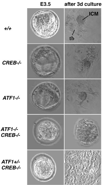
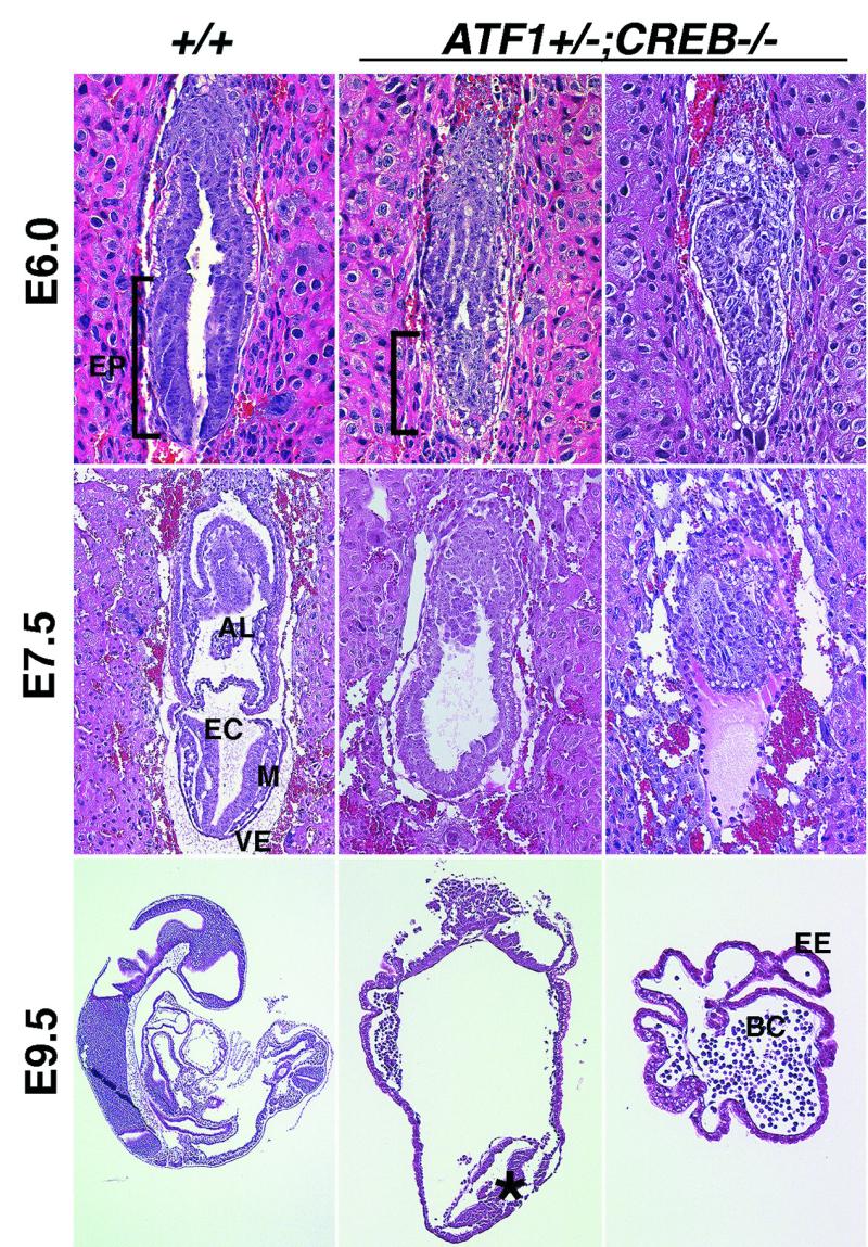
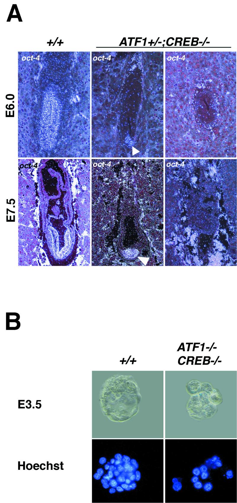
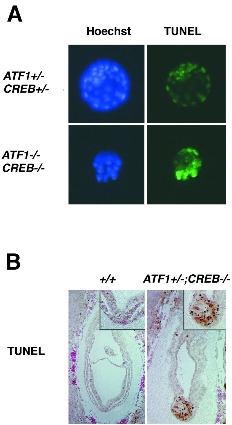
Activating transcription factor 1 (ATF1), CREB, and the cyclic AMP (cAMP) response element modulatory protein (CREM), which constitute a subfamily of the basic leucine zipper transcription factors, activate gene expression by binding as homo- or heterodimers to the cAMP response element in regulatory regions of target genes. To investigate the function of ATF1 in vivo, we inactivated the corresponding gene by homologous recombination. In contrast to CREB-deficient mice, which suffer from perinatal lethality, mice lacking ATF1 do not exhibit any discernible phenotypic abnormalities. Since ATF1 and CREB but not CREM are strongly coexpressed during early mouse development, we generated mice deficient for both CREB and ATF1. ATF1(-/-) CREB(-/-) embryos die before implantation due to developmental arrest. ATF1(+/-) CREB(-/-) embryos display a phenotype of embryonic lethality around embryonic day 9.5 due to massive apoptosis. These results indicate that CREB and ATF1 act in concert to mediate signals essential for maintaining cell viability during early embryonic development.


Figures

FIG. 1.
FIG. 1.
Immunohistochemical analysis revealed overlapping expression of ATF1 and CREB during early embryogenesis. At E3.5 ATF1 could be detected in the whole blastocyst, whereas CREB was mainly located in the ICM cells. During further embryogenesis at E6.0 and E7.5, ATF1 is expressed in all embryonic, extraembryonic, and trophectoderm-derived cells. CREB was found only in epiblast cells and lineages deriving thereof. A clear border can be seen between extraembryonic parts and the epiblast (arrowheads). In adult testis, the expression pattern of both proteins is very distinct. ATF1 is highly expressed in spermatocytes of the pachytene stage, whereas CREB protein is found only in Sertoli cells (arrows).
FIG. 2.
FIG. 2.
Inactivation of the mouse ATF1 gene by gene targeting. (A) Shown is part of the ATF1 locus with exons 3 to 7 (black boxes) encoding portions of the kinase-inducible domain (exon 3) and all of the leucine zipper domain (exon 7) (top). Targeting construct containing a 3.8-kb 5′ homology and a 1.6-kb 3′ homology fragment inserted into the pHM3 vector is shown (middle). The targeted ATF1 gene is shown at bottom. lacZ-neo, β-galactosidase/neomycin-resistance cassette; Bg, BglII; E, EcoRI. (B) Top, PCR genotyping of genomic DNA isolated from embryos of heterozygous intercrosses. Detection of the wild-type allele corresponds to a 260-bp fragment, and the mutant allele corresponds to a 420-bp fragment. Bottom, Western blot analysis of nucleus extracts of kidney, liver, and testis shows clear loss of ATF1 in tissues of ATF1−/− mice. (C) Whole-mount β-galactosidase stainings of ATF1+/− and CREB+/− embryos. LacZ staining confirms the expression pattern of ATF1 and CREB revealed by immunohistochemistry (Fig. 1).
FIG. 3.
FIG. 3.
Functional disruption of both CREB and ATF1 results in preimplantation defects. Embryos of different genotypes were isolated at E3.5. ATF1+/− CREB−/− and ATF1−/− CREB−/− embryos show a morula-like structure, while wild-type and single-knockout embryos are comprised of trophectoderm surrounding ICM and blastocoel. Outgrowths after 4 days of in vitro culture of wild-type, ATF1−/−, and CREB−/− blastocysts develop a monolayer of trophoblast cells with the ICM cells on top. No outgrowth of ATF1−/− CREB−/− embryos occurs after 4 days in culture. Embryos seem to arrest in development; cells appear necrotic and are still surrounded by the zona pellucida. ATF1+/− CREB−/− embryos hatch from the zona pellucida; a trophoblast monolayer is present, but no obvious ICM can be seen. tb, trophoblast monolayer. 3d, 3 days.
FIG. 4.
FIG. 4.
ATF1+/− CREB−/− embryos exhibit severe embryonic disorganization. Sections of hematoxylin-and-eosin-stained wild-type and ATF1+/− CREB−/− embryos at embryonic stages E6.0, E7.5, and E9.5 are shown. Two types of ATF1+/− CREB−/− embryos could be isolated. One group showed a strong reduction and malformation of the embryonic region, whereas the other one was comprised only of extraembryonic ectoderm lacking the embryonic part completely. At E6.0 ATF1+/− CREB−/− embryos fail to form the pseudostratified columnar epithelium of the epiblast. At E7.5, gastrulation is almost complete and the three germ layers have developed from the epiblast. In ATF1+/− CREB−/− embryos, germ layers cannot be distinguished and extraembryonic mesoderm-derived structures like amnion, allantois, and chorion are missing. E9.5 embryos of this genotype fail to develop any structural organization compared to wild-type embryos of the same age. Some of the ATF1+/− CREB−/− embryos develop, instead of a structured embryo, a group of mesoderm-like cells (star) at the distal end of the conceptus. Others comprise only blood cells surrounded by extraembryonic ectoderm. AL, allantois; EC, embryonic ectoderm; EP, epiblast; M, mesoderm; VE, visceral endoderm; EE, extraembryonic ectoderm; and BC, blood cells.
FIG. 5.
FIG. 5.
Number of totipotent cells in ATF1−/− CREB−/− embryos and of pluripotent cells in ATF1+/− CREB−/− embryos is strongly reduced. (A) oct-4 expression in wild-type and ATF1+/− CREB−/− embryos at E6.0 and E7.5 detected by in situ hybridization. Pluripotent cells of the epiblast appear positive for Oct-4. In contrast to the wild type, ATF1+/− CREB−/− embryos develop a strongly reduced embryonic part with fewer epiblast cells or lack pluripotent cells completely. (B) Hoechst staining reveals that ATF1−/− CREB−/− embryos are comprised of fewer blastomeres than are their wild-type littermates.
FIG. 6.
FIG. 6.
Both ATF1 and CREB are important for cellular survival during early development. In situ cell death detection shows an increasing number of dying cells in ATF1−/− CREB−/− as well as in ATF1+/− CREB−/− embryos but at different developmental stages. (A) After 1 day of culture, control blastocysts do not show any TUNEL-labeled cells, whereas almost all cells of ATF1−/− CREB−/− embryos, isolated at E3.5, seem to be dying. (B) At E7.5, ATF1+/− CREB−/− mutant embryos show, compared to the wild types, intense TUNEL staining in the distal part of the embryo overlapping the oct-4-expressing region.

References

    1. Ang, S. L., and J. Rossant. 1994. HNF-3 beta is essential for node and notochord formation in mouse development. Cell 78:561-574. - PubMed
    1. Barnes, J. D., J. L. Crosby, C. M. Jones, C. V. Wright, and B. L. Hogan. 1994. Embryonic expression of Lim-1, the mouse homolog of Xenopus Xlim-1, suggests a role in lateral mesoderm differentiation and neurogenesis. Dev. Biol. 161:168-178. - PubMed
    1. Blendy, J. A., K. H. Kaestner, G. F. Weinbauer, E. Nieschlag, and G. Schutz. 1996. Severe impairment of spermatogenesis in mice lacking the CREM gene. Nature 380:162-165. - PubMed
    1. Bonni, A., A. Brunet, A. E. West, S. R. Datta, M. A. Takasu, and M. E. Greenberg. 1999. Cell survival promoted by the Ras-MAPK signaling pathway by transcription-dependent and -independent mechanisms. Science 286:1358-1362. - PubMed
    1. Bosilevac, J. M., R. J. Olsen, J. A. Bridge, and S. H. Hinrichs. 1999. Tumor cell viability in clear cell sarcoma requires DNA binding activity of the EWS/ATF1 fusion protein. J. Biol. Chem. 274:34811-34818. - PubMed

Publication types

MeSH terms

Substances

LinkOut - more resources