. 2002 Mar;22(6):1947-60.
doi: 10.1128/MCB.22.6.1947-1960.2002.

Diverse effects of mutations in exon II of the von Hippel-Lindau (VHL) tumor suppressor gene on the interaction of pVHL with the cytosolic chaperonin and pVHL-dependent ubiquitin ligase activity

Affiliations

Diverse effects of mutations in exon II of the von Hippel-Lindau (VHL) tumor suppressor gene on the interaction of pVHL with the cytosolic chaperonin and pVHL-dependent ubiquitin ligase activity

William J Hansen et al. Mol Cell Biol. 2002 Mar.

Abstract

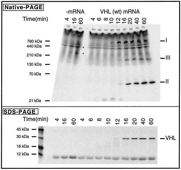
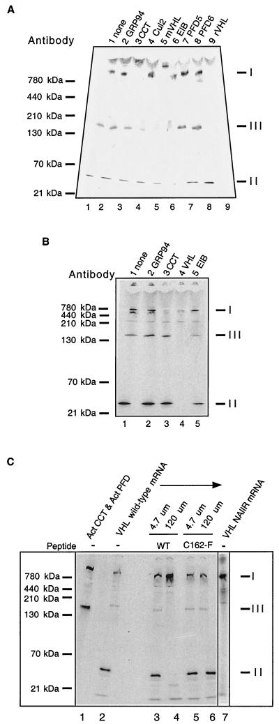
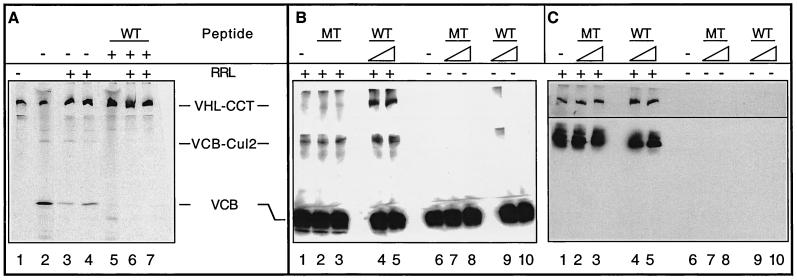
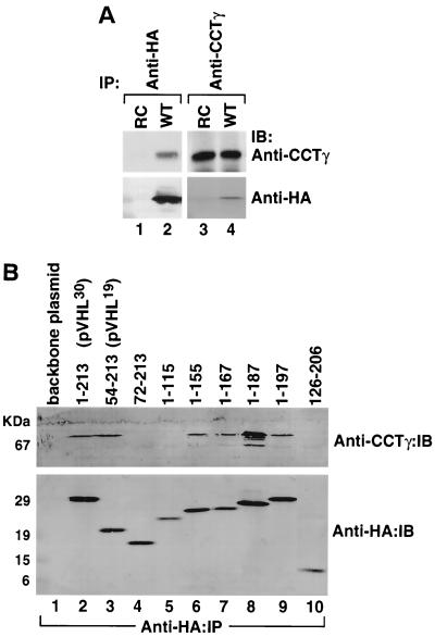
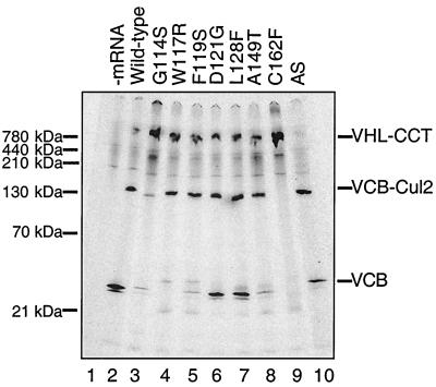
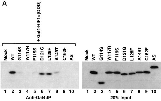
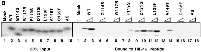
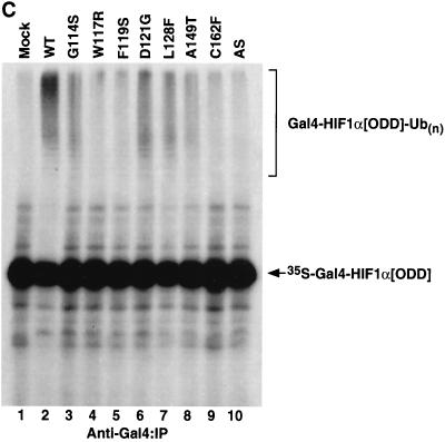
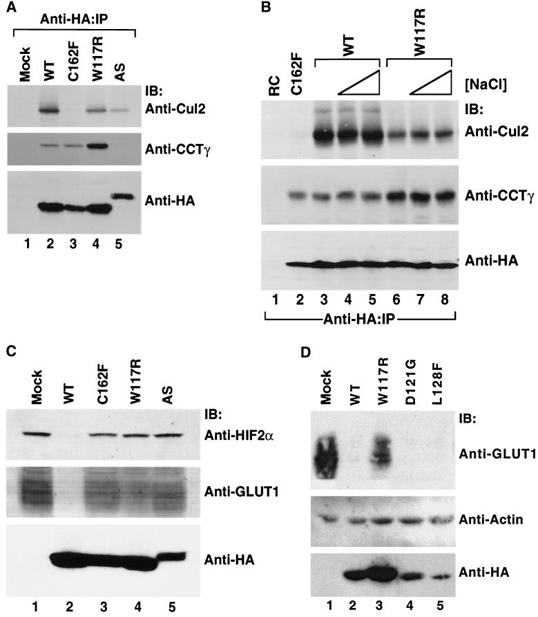
We examined the biogenesis of the von Hippel-Lindau (VHL) tumor suppressor protein (pVHL) in vitro and in vivo. pVHL formed a complex with the cytosolic chaperonin containing TCP-1 (CCT or TRiC) en route to assembly with elongin B/C and the subsequent formation of the VCB-Cul2 ubiquitin ligase. Blocking the interaction of pVHL with elongin B/C resulted in accumulation of pVHL within the CCT complex. pVHL present in purified VHL-CCT complexes, when added to rabbit reticulocyte lysate, proceeded to form VCB and VCB-Cul2. Thus, CCT likely functions, at least in part, by retaining VHL chains pending the availability of elongin B/C for final folding and/or assembly. Tumor-associated mutations within exon II of the VHL syndrome had diverse effects upon the stability and/or function of pVHL-containing complexes. First, a pVHL mutant lacking the entire region encoded by exon II did not bind to CCT and yet could still assemble into complexes with elongin B/C and elongin B/C-Cul2. Second, a number of tumor-derived missense mutations in exon II did not decrease CCT binding, and most had no detectable effect upon VCB-Cul2 assembly. Many exon II mutants, however, were found to be defective in the binding to and subsequent ubiquitination of hypoxia-inducible factor 1alpha (HIF-1alpha), a substrate of the VCB-Cul2 ubiquitin ligase. We conclude that the selection pressure to mutate VHL exon II during tumorigenesis does not relate to loss of CCT binding but may reflect quantitative or qualitative defects in HIF binding and/or in pVHL-dependent ubiquitin ligase activity.


Figures

FIG. 1.
FIG. 1.
Order of events in the assembly pathway of the VCB and VCB-Cul2 complexes. (Top) VHL mRNA was translated in the RRL in the presence of [35S]methionine for 4 min, and then inhibitors of initiation of translation were added, followed by continued incubation. Equal aliquots were removed at the times indicated after the beginning of the reaction and analyzed by native-PAGE. A fluorogram of the gel is shown. The migration positions of different pVHL-containing complexes are indicated on the right as I, II, and III (based on their apparent order of appearance). Native molecular mass standards are shown on the left. A control translation reaction in which no mRNA was added (−mRNA) was analyzed on the left. The position of migration of species not dependent upon VHL mRNA (i.e., which was present in the no-added mRNA column) is indicated by an asterisk. (Bottom) Aliquots of the same samples examined in the top panel were analyzed by SDS-PAGE. A fluorogram of the gel is shown. The migration positions of molecular mass standards are shown on the left.
FIG. 2.
FIG. 2.
Identification of pVHL-containing complexes formed during in vitro translation of VHL mRNA. (A) Antibody-induced gel shifts of pVHL-containing complexes. [35S]methionine-labeled pVHL-containing complexes were generated by in vitro translation of VHL mRNA in the RRL. Cycloheximide then was added to a final concentration of 0.5 mM, and equal aliquots of the reaction products were incubated at 4°C for 30 min with the following antibodies: lane 1, noantibody; lane 2, rat monoclonal anti-GRP94 (negative control); lane 3, rat monoclonal anti-CCT-1α; lane 4, rabbit polyclonal anti-Cul2; lane 5, mouse monoclonal anti-pVHL; lane 6, mouse monoclonal and goat anti-elongin B; lane 7, rabbit anti-prefoldin subunit 5; lane 8, rabbit anti-prefoldin subunit 6; and lane 9, rabbit anti-pVHL. The reaction products were then analyzed by native-PAGE. A fluorogram of the gel is shown. The migration positions of the pVHL complexes are indicated at the right (indicated by I, II, and III), and those of native molecular mass standards are shown on the left. (B) Binding of pVHL-containing complexes to immobilized antibodies. pVHL-containing complexes similar to those in panel A were generated by in vitro translation of VHL mRNA in the RRL. Equal aliquots of the reaction products were incubated at 4°C for 30 min with the following antibodies immobilized on a mixture of protein A- and protein G-Sepharose: lane 1, no antibody; lane 2, rat monoclonal anti-GRP94 (negative control); lane 3, rat monoclonal anti-CCT-1α; lane 4, mouse monoclonal anti-VHL and rabbit VHL antibodies; and lane 5, mouse monoclonal and goat anti-elongin B. Proteins remaining in the unbound fraction were then analyzed by native-PAGE. A fluorogram of the gel is shown. The migration positions of the pVHL complexes are indicated on the right (by I, II, and III), and those of the native molecular mass standards are shown on the left. (C) A peptide derived from pVHL, known to interact with elongin C, blocks the formation of complex II and III and results in the accumulation of VHL with CCT (complex I). VHL mRNA was translated in the presence of [35S]methionine in the RRL for 60 min in the absence or presence of two different synthetic pVHL peptides (at the concentrations indicated at the top). The first peptide was derived from wild-type pVHL (amino acids 157 to 172) and is known to bind to elongin C (WT), and the second was derived from a mutant form of pVHL that fails to interact with elongin C (C162F). The lane on the far right contains the products of a translation reaction programmed with VHL mRNA encoding a mutant that contains an insertion of the sequence NAIIRS in the elongin C binding region of pVHL. A fluorogram of the native gel is shown. The migration positions of native molecular mass standards are indicated at the far left. For reference purposes, a reaction wherein newly synthesized actin bound to both CCT and prefoldin is included in the first lane. The position of migration of the pVHL complexes is indicated on the right (I, II, and III).
FIG. 3.
FIG. 3.
A role for CCT in the assembly and disassembly of VCB. (A) Generation of VCB from purified VHL-CCT complexes. VHL-CCT complexes assembled in RRL were isolated by gel filtration chromatography and analyzed by native-PAGE (lane 1). To mark the position of migration of the VCB and VCB-Cul2 complexes, a sample of the unfractionated RRL reaction also was analyzed (lane 2). Purified VHL-CCT was incubated in the presence of RRL and an ATP-regeneration system (lanes 3 and 4), which resulted in the appearance of both VCB and VCB-Cul2. The migration position of VHL-CCT is marked by pVHL synthesized in the presence of the elongin C binding peptide (lane 5). When purified VHL-CCT was incubated in RRL in the presence of the elongin C binding peptide, VCB and VCB-Cul2 formation now was blocked (lanes 6 and 7). (B and C) pVHL present in the VCB complex rebinds to CCT upon VCB disassembly. (B) Recombinant VCB (see Materials and Methods) was added to rabbit reticulocyte lysate (+RRL, lanes 1 to 5) and then incubated in the absence (lane 1) or presence of either the pVHL peptide (WT, lanes 4 and 5) or the mutant pVHL peptide (MT, lanes 2 and 3) described in Fig. 2C (either 120 or 600 μM as indicated by the triangles). As a control, the recombinant VCB was incubated in the absence (lane 6) or presence of either wild type (WT, lanes 9 and 10) or mutant (MT, lanes 7 and 8) peptide in the absence of RRL (−RRL, lanes 6 to 10). After 60 min of incubation, the reaction products were analyzed by native-PAGE. The proteins in the gel were denatured, transferred to nitrocellulose, and analyzed in panel B by Western blotting with a monoclonal antibody specific for pVHL. The positions of the pVHL-containing complexes are indicated at the left. (C) The membrane from the blot shown in panel B was stripped and reprobed with anti-CCT-1α (top) or anti-Cul2 antibodies (bottom). The lane designations are the same as for panel B.
FIG. 4.
FIG. 4.
(A) Interactions of wild-type and mutant pVHL with the cytosolic chaperonin in vivo. RCC 786-O cells (lacking endogenous pVHL) transfected with and stably expressing HA-tagged wild-type pVHL (WT) or cells transfected with empty plasmid (RC) were lysed and used for immunoprecipitation reactions (IP) employing an anti-HA antibody (lanes 1 and 2) or an anti-CCTγ antibody (lanes 3 and 4). The resultant pVHL immunoprecipitates were resolved by SDS-PAGE, transferred onto a polyvinylidene difluoride membrane, and then immunoblotted (IB) with anti-CCTγ (top panels) or anti-HA (bottom panels) antibodies. (B) The region encompassing amino acids 54 to 155 of pVHL contributes to the binding of CCT. RCC 786-O cells expressing HA-tagged versions of either wild-type pVHL or a series of pVHL deletion mutants were lysed and used for immunoprecipitation reactions with the anti-HA antibody. The resultant pVHL immunoprecipitates were resolved by SDS-PAGE, transferred to a polyvinylidene difluoride membrane, and then analyzed for the presence of both the cytosolic chaperonin (Anti-CCTγ:IB) and HA-pVHL proteins (Anti-HA:IB) via Western blotting. The amino acids included in each pVHL polypeptide are listed at the top. Lane 1 represents cells transfected with empty plasmid. p30 represents the full-length pVHL that was initiated at the first methionine codon, while p19 represents a naturally occurring pVHL that was initiated at the second methionine codon.
FIG. 5.
FIG. 5.
Effects of mutations in VHL exon II on the binding of pVHL to CCT and the assembly of VCB/VCB-Cul2. mRNAs encoding either wild-type or various mutant forms of pVHL were translated in the RRL in the presence of [35S]methionine for 45 min, and aliquots of the reactions were removed and analyzed by native-PAGE. The migration positions of the pVHL-containing complexes are indicated on the right, and the positions of native molecular mass standards are shown on the left. A control translation reaction (−mRNA) was analyzed in lane 1. An exon III mutant, C162F, which does not form VCB and VCB-Cul2 was analyzed as a negative control in lane 9. AS is the naturally occurring alternatively spliced mutant in which all of exon II is deleted. The particular pVHL polypeptide synthesized in each reaction is indicated at the top of the panel.
FIG. 6.
FIG. 6.
Diverse effects of mutations in exon II of VHL on ability of VCB-Cul2 to interact with the HIF-1α sequence implicated in oxygen-dependent proteolysis. (A) Wild-type and mutant VHL mRNAs were translated in the RRL in the presence of [35S]methionine. In a separate reaction, Gal4-HIF-1α[ODD] mRNA was translated (in the absence of [35S]methionine). Equal portions of the two reactions were mixed together in order to allow the 35S-labeled VCB-Cul2 to interact with the Gal4-HIF-1α[ODD] substrate. After a 30-min incubation, the Gal4-HIF-1α[ODD] was captured by using an anti-Gal4 antibody bound to protein A-Sepharose. The amount of 35S-labeled pVHL coprecipitating was determined by SDS-PAGE and fluorography. In the right panel, 20% of the input radiolabeled pVHL proteins used in the analysis are shown. In the left panel, the radiolabeled pVHL proteins immunoprecipitated by the anti-Gal4 antibodies are shown. The particular pVHL polypeptide synthesized in each reaction is indicated at the top of the panel. Note that the G114S mutant pVHL (lane 3) was observed to migrate faster than the wild-type (lane 2) and that the exon II deletion mutant (lane 10) migrated slightly slower than the wild-type protein under these conditions during SDS-PAGE in Tris-glycine buffer. The reason for this anomalous migration is not known. These anomalies were not observed when Tris-Tricine gels were used. (B) Binding of pVHL encoded by exon II mutants to a hydroxylated HIF-derived peptide. The indicated wild-type and mutant VHL mRNAs were translated in RRL in the presence of [35S]methionine. Then, 5 or 10 μl of radiolabeled translation products, as indicated by the triangles, was incubated with 0.1 μg of an immobilized biotin-conjugated hydroxylated HIF-derived peptide (a substrate for the VCB-Cul2 complex) (see Materials and Methods) to allow 35S-labeled VCB-Cul2 to interact with the hydroxylated peptide. After a 1-h incubation at 4°C, the amount of bound 35S-labeled pVHL was determined by SDS-PAGE and fluorography (right panel). In the left panel, 20% of the input radiolabeled pVHL proteins used in the analysis is shown. “Mock” indicates a reaction that was programmed with empty plasmid. (C) The ability of VHL exon II mutants to stimulate the ubiquitination of HIF-1α correlates well with the binding of the pVHL variants to HIF-1α. In an experiment similar to that shown in panels A and B, Gal4-HIF-1α[ODD] was synthesized in RRL, this time in the presence of [35S]methionine. In parallel, the wild type and the different pVHL mutants were translated separately in the absence of radiolabel. Portions of the translation reactions (4 μl of 35S-labeled Gal4-HIF-1α[ODD] and 2 μl of each unlabeled pVHL) were mixed together, along with an in vitro system capable of ubiquitin conjugation (see Materials and Methods). The Gal4-HIF-1α[ODD] was isolated by immunoprecipitation with anti-Gal4 antibodies, and the resultant immunoprecipitates were analyzed by SDS-PAGE and fluorography. The particular pVHL polypeptide synthesized in each reaction is indicated at the top of the panel. Indicated near the top of the gel is the position of ubiquitin-conjugated Gal4-HIF-1α[ODD] [UB(n)]. “Mock” indicates a reaction that was programmed with empty plasmid.
FIG. 6.
FIG. 6.
Diverse effects of mutations in exon II of VHL on ability of VCB-Cul2 to interact with the HIF-1α sequence implicated in oxygen-dependent proteolysis. (A) Wild-type and mutant VHL mRNAs were translated in the RRL in the presence of [35S]methionine. In a separate reaction, Gal4-HIF-1α[ODD] mRNA was translated (in the absence of [35S]methionine). Equal portions of the two reactions were mixed together in order to allow the 35S-labeled VCB-Cul2 to interact with the Gal4-HIF-1α[ODD] substrate. After a 30-min incubation, the Gal4-HIF-1α[ODD] was captured by using an anti-Gal4 antibody bound to protein A-Sepharose. The amount of 35S-labeled pVHL coprecipitating was determined by SDS-PAGE and fluorography. In the right panel, 20% of the input radiolabeled pVHL proteins used in the analysis are shown. In the left panel, the radiolabeled pVHL proteins immunoprecipitated by the anti-Gal4 antibodies are shown. The particular pVHL polypeptide synthesized in each reaction is indicated at the top of the panel. Note that the G114S mutant pVHL (lane 3) was observed to migrate faster than the wild-type (lane 2) and that the exon II deletion mutant (lane 10) migrated slightly slower than the wild-type protein under these conditions during SDS-PAGE in Tris-glycine buffer. The reason for this anomalous migration is not known. These anomalies were not observed when Tris-Tricine gels were used. (B) Binding of pVHL encoded by exon II mutants to a hydroxylated HIF-derived peptide. The indicated wild-type and mutant VHL mRNAs were translated in RRL in the presence of [35S]methionine. Then, 5 or 10 μl of radiolabeled translation products, as indicated by the triangles, was incubated with 0.1 μg of an immobilized biotin-conjugated hydroxylated HIF-derived peptide (a substrate for the VCB-Cul2 complex) (see Materials and Methods) to allow 35S-labeled VCB-Cul2 to interact with the hydroxylated peptide. After a 1-h incubation at 4°C, the amount of bound 35S-labeled pVHL was determined by SDS-PAGE and fluorography (right panel). In the left panel, 20% of the input radiolabeled pVHL proteins used in the analysis is shown. “Mock” indicates a reaction that was programmed with empty plasmid. (C) The ability of VHL exon II mutants to stimulate the ubiquitination of HIF-1α correlates well with the binding of the pVHL variants to HIF-1α. In an experiment similar to that shown in panels A and B, Gal4-HIF-1α[ODD] was synthesized in RRL, this time in the presence of [35S]methionine. In parallel, the wild type and the different pVHL mutants were translated separately in the absence of radiolabel. Portions of the translation reactions (4 μl of 35S-labeled Gal4-HIF-1α[ODD] and 2 μl of each unlabeled pVHL) were mixed together, along with an in vitro system capable of ubiquitin conjugation (see Materials and Methods). The Gal4-HIF-1α[ODD] was isolated by immunoprecipitation with anti-Gal4 antibodies, and the resultant immunoprecipitates were analyzed by SDS-PAGE and fluorography. The particular pVHL polypeptide synthesized in each reaction is indicated at the top of the panel. Indicated near the top of the gel is the position of ubiquitin-conjugated Gal4-HIF-1α[ODD] [UB(n)]. “Mock” indicates a reaction that was programmed with empty plasmid.
FIG. 7.
FIG. 7.
Stability and function of wild-type and representative exon II mutant pVHL polypeptide complexes with CCT and elongin B/C-Cul2 formed in vivo. (A) Effects of mutations in VHL exon II upon pVHL-CCT and VCB assembly. RCC 786-O cells expressing empty plasmid (Mock) or HA-tagged versions of wild-type or mutant pVHL were lysed and then immunoprecipitated with the anti-HA antibody (Anti-HA-IP). Resultant immunoprecipitates were resolved by SDS-PAGE and then Western blotted with anti-Cul2 (top panel), anti-CCTγ (middle panel), and anti-HA (bottom panel) antibodies. The particular pVHL polypeptide synthesized is indicated at the top of the panel. (B) Effects on stability of pVHL-CCT and VCB-Cul2 complexes. RCC 786-O cells expressing HA-tagged wild-type pVHL (WT) (lanes 3 to 5) or an exon II pVHL point mutant (W117R) (lanes 6 to 8) were lysed and immunoprecipitated with the anti-HA antibody under increasing NaCl concentrations (125 mM [lanes 1, 2, 3, and 6], 500 mM [lanes 4 and 7], and 900 mM [lanes 5 and 8]). Lysates from cells transfected with plasmid alone (RC) (lane 1) and the HA-tagged exon III mutant (C162F) (lane 2) were immunoprecipitated with the anti-HA antibody under the lowest NaCl stringency condition. Proteins present within the immunoprecipitates were resolved by SDS-PAGE, transferred to nitrocellulose, and analyzed via Western blotting (IB) with the anti-Cul2 (top panel), anti-CCTγ (middle panel), and anti-HA (bottom panel) antibodies. (C) Exon II mutants that do not bind to HIF in the in vitro assays are unable to suppress expression of a HIF 2α regulated gene product, GLUT1. In parallel, the same cell lysates analyzed in B were examined for their expression of HIF-2α and GLUT1, a gene product whose expression is regulated by the HIF-2α transcription factor. Whole-cellextracts were prepared from the cells described above, and the proteins (200 μg) were resolved by SDS-PAGE and then Western blotted (IB) with anti-HIF-2α (top panel), anti-GLUT1 (middle panel), or anti-HA (bottom panel) antibodies. The GLUT1 protein likely appears as a rather diffuse band due to heterogeneity in its glycosylation and because of its many (>10) transmembrane segments (35, 57). (D) Exon II mutants that do bind to HIF in the in vitro assays are able to suppress expression of an HIF-2α-regulated gene product. RCC 786-O cells were infected with retroviral vectors encoding HA-tagged versions of the wild type or the indicated pVHL mutants. Whole-cell extracts were prepared, and proteins (200 μg) were resolved by SDS-PAGE and Western blotted (IB) with anti-GLUT1 (top panel), anti-actin (middle panel), or anti-HA (bottom panels) antibodies.

Similar articles

Cited by

References

    1. Blankenship, C., J. G. Naglich, J. M. Whaley, B. Seizinger, and N. Kley. 1999. Alternate choice of initiation codon produces a biologically active product of the von Hippel Lindau gene with tumor suppressor activity. Oncogene 18:1529-1535. - PubMed
    1. Bonicalzi, M. E., I. Groulx, N. de Paulsen, and S. Lee. 2001. Role of exon 2-encoded β-domain of the von Hippel-Lindau tumor suppressor protein. J. Biol. Chem. 276:1407-1416. - PubMed
    1. Botuyan, M. V., C. M. Koth, G. Mer, A. Chakrabartty, J. W. Conaway, R. C. Conaway, A. M. Edwards, C. H. Arrowsmith, and W. J. Chazin. 1999. Binding of elongin A or a von Hippel-Lindau peptide stabilizes the structure of yeast elongin C. Proc. Natl. Acad. Sci. USA 96:9033-9038. - PMC - PubMed
    1. Cockman, M. E., N. Masson, D. R. Mole, P. Jaakkola, G. W. Chang, S. C. Clifford, E. R. Maher, C. W. Pugh, P. J. Ratcliffe, and P. H. Maxwell. 2000. Hypoxia inducible factor-alpha binding and ubiquitylation by the von Hippel-Lindau tumor suppressor protein. J. Biol. Chem. 275:25733-25741. - PubMed
    1. Cowan, N. J., and S. A. Lewis. 1999. A chaperone with a hydrophilic surface. Nat. Struct. Biol. 6:990-991. - PubMed

Publication types

MeSH terms