. 2002 Mar 1;21(5):876-84.
doi: 10.1093/emboj/21.5.876.

The morphogenic linker peptide of HBV capsid protein forms a mobile array on the interior surface

Affiliations

The morphogenic linker peptide of HBV capsid protein forms a mobile array on the interior surface

Norman R Watts et al. EMBO J. .

Abstract

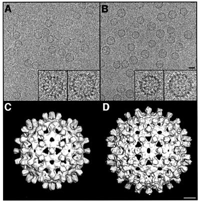
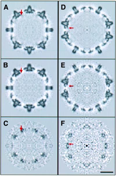
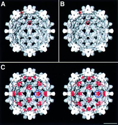
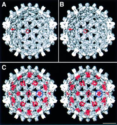
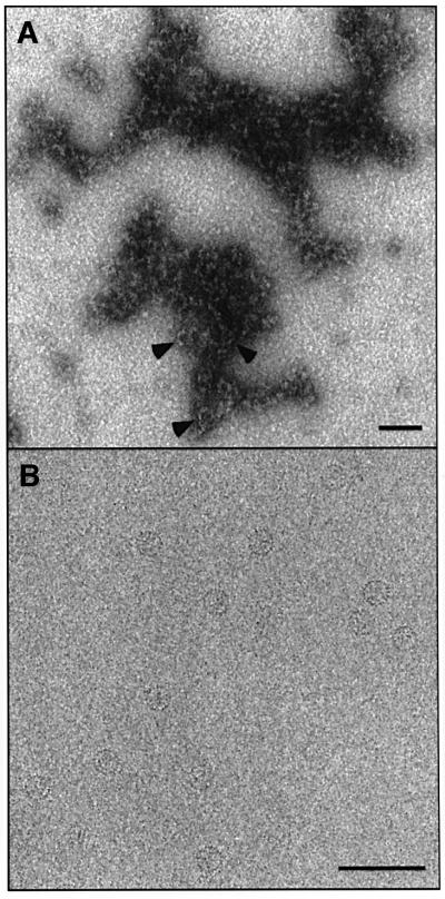
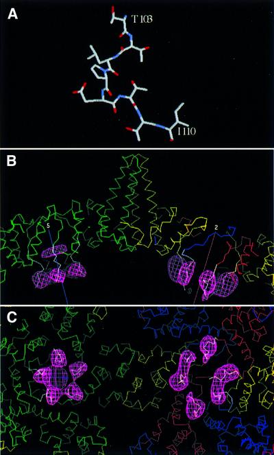
Many capsid proteins have peptides that influence their assembly. In hepatitis B virus capsid protein, the peptide STLPETTVV, linking the shell-forming 'core' domain and the nucleic acid-binding 'protamine' domain, has such a role. We have studied its morphogenic properties by permuting its sequence, substituting it with an extraneous peptide, deleting it to directly fuse the core and protamine domains and assembling core domain dimers with added linker peptides. The peptide was found to be necessary for the assembly of protamine domain-containing capsids, although its size-determining effect tolerates some modifications. Although largely invisible in a capsid crystal structure, we could visualize linker peptides by cryo-EM difference imaging: they emerge on the inner surface and extend from the capsid protein dimer interface towards the adjacent symmetry axis. A closely sequence-similar peptide in cellobiose dehydrogenase, which has an extended conformation, offers a plausible prototype. We propose that linker peptides are attached to the capsid inner surface as hinged struts, forming a mobile array, an arrangement with implications for morphogenesis and the management of encapsidated nucleic acid.


Figures

None
Fig. 1. (A and B) Cryo-electron micrographs of Cp140 and Cp149 capsids. Bar = 250 Å. In each case, examples of a T = 3 and a T = 4 capsid are shown at higher magnification (×3) in the inserts. (C and D) Surface renderings of the outside of Cp140 capsids: (C) T = 3; (D) T = 4. Bar = 50 Å.
None
Fig. 2. Central sections through T = 3 capsids viewed along a 2-fold axis. The red arrows mark one of the locations at which extra density is present in the Cp149 capsid (A) but not the Cp140 capsid (B). This density is also present at other quasi-equivalent, independently calculated, sites on the icosahedral surface lattice. These densities are more conspicuous in the corresponding section through the difference map (C). Bar = 100 Å. (DF) show the corresponding sections for T = 4 capsids.
None
Fig. 3. (A and B) Interior surfaces of the T = 3 capsids of Cp140 and Cp149. Additional densities present on Cp149 are marked with a red arrow and ring. (C) Stereo rendering of the Cp140 capsid with the difference densities juxtaposed and contoured at the same level (pink). Difference density at the 5-fold is marked with a blue ring. Bar = 100 Å.
None
Fig. 4. (A and B) Interior surfaces of the T = 4 capsids of Cp140 and Cp149. Additional densities present on Cp149 are marked with a red arrow and a ring. (C) Stereo rendering of the Cp140 capsid with the difference densities juxtaposed and contoured at the same level (pink). Difference density at the 5-fold is marked with a blue ring. Bar = 100 Å.
None
Fig. 5. Effects on assembly of substituting the last seven residues of linker peptide. (A) Cp142ETVPVLT, permuting the native sequence, substitutes a heptapeptide of the same size, net charge and polarity. Under conditions conducive to capsid assembly, most of the material forms aggregates, shown here by negative staining. In places, its appearance suggests a condensate of partially formed capsids. Very few complete capsids are present (arrowheads). (B) Substitution with the N-terminal heptapeptide. This construct, Cp142MDIDPYK, assembled normally, producing almost exclusively T = 4 particles as illustrated here by cryo-EM. Bar = 1000 Å.
None
Fig. 6. Modeling a peptide structure into linker-associated densities visualized by cryo-EM difference imaging. Residues 103–110 of cellobiose dehydrogenase (Hallberg et al., 2000) are almost completely identical to the linker sequences (A). In (B), this peptide is shown (white α-carbon trace) docked into the difference density (pink mesh) from the T = 4 capsids. Quasi-equivalent core domain subunits are shown in green, yellow, red and blue. Five-fold (at left) and 2-fold (at right) symmetry axes are marked. In (C), the view is from the inside of the capsid. We propose that a semi-flexible hinge allows the linker to pivot, so that it serves as a spacer that prevents the protamine domains from intruding too closely into inter-core domain contacts, and also confers mobility on the protamine domains’ interactions with nucleic acid.
None
Fig. 7. (A) Quasi-atomic model of the T = 3 HBV Cp142 capsid. The model was created by docking dimers from the crystal structure of the T = 4 capsid into a cryo-EM map of the T = 3 capsid. (B and D) Central sections through quasi-atomic models of the T = 3 (B) and T = 4 (D) capsids. Cp142 capsids, restricted to 10 Å resolution, closely resemble the corresponding sections through the cryo-EM density maps of Cp149 and Cp140 (cf. Figures 2A, B, D and E). Corresponding sections through quasi-atomic models of the icosahedrally coordinated linker peptide (represented by the cellobiose dehydrogenase peptide, appropriately docked) are shown in (C) and (E) (cf. Figure 2C and F). Bar = 100 Å.

References

    1. Baker T.S. and Cheng,R.H. (1996) A model-based approach for determining orientations of biological macromolecules imaged by cryoelectron microscopy. J. Struct. Biol., 116, 120–130. - PubMed
    1. Bartenschlager R. and Schaller,H. (1992) Hepadnaviral assembly is initiated by polymerase binding to the encapsidation signal in the viral RNA genome. EMBO J., 11, 3413–3420. - PMC - PubMed
    1. Beames B. and Lanford,R.E. (1993) Carboxy-terminal truncations of the HBV core protein affect capsid formation and the apparent size of encapsidated HBV RNA. Virology, 194, 597–607. - PubMed
    1. Belnap D.M., Kumar,A., Folk,J.T., Smith,T.J. and Baker,T.S. (1999) Low-resolution density maps from atomic models: how stepping ‘back’ can be a step ‘forward’. J. Struct. Biol., 125, 166–175. - PubMed
    1. Birnbaum F. and Nassal,M. (1990) Hepatitis B virus nucleocapsid assembly: primary structure requirements in the core protein. J. Virol., 64, 3319–3330. - PMC - PubMed

Publication types

MeSH terms

Substances