Comparative Study
. 2002 Mar 1;21(5):885-97.
doi: 10.1093/emboj/21.5.885.

The rhesus rotavirus VP4 sialic acid binding domain has a galectin fold with a novel carbohydrate binding site

Affiliations
Comparative Study

The rhesus rotavirus VP4 sialic acid binding domain has a galectin fold with a novel carbohydrate binding site

Philip R Dormitzer et al. EMBO J. .

Abstract

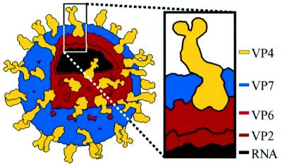
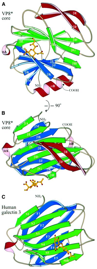
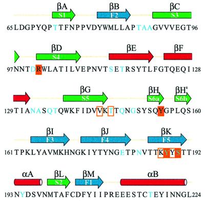
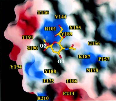
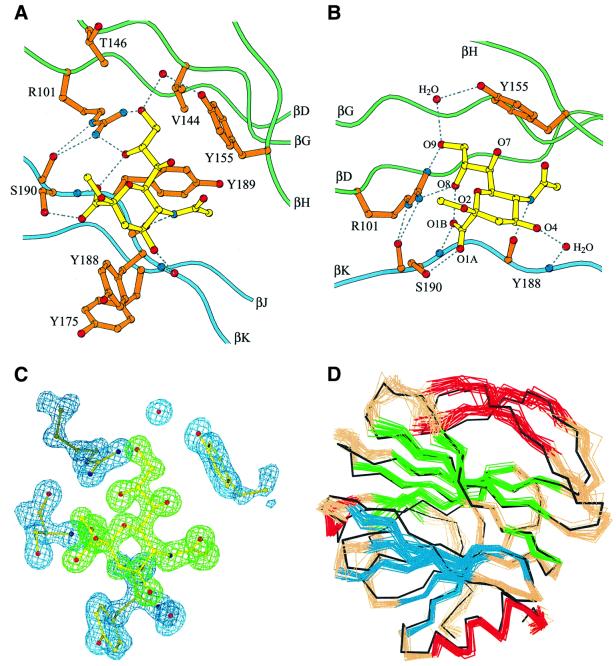
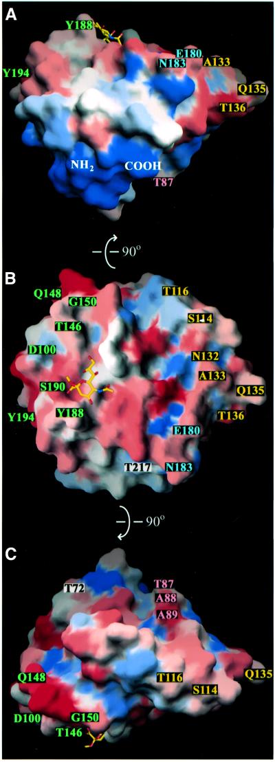
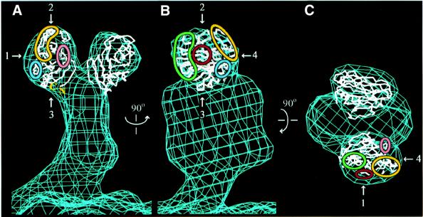
Cell attachment and membrane penetration are functions of the rotavirus outer capsid spike protein, VP4. An activating tryptic cleavage of VP4 produces the N-terminal fragment, VP8*, which is the viral hemagglutinin and an important target of neutralizing antibodies. We have determined, by X-ray crystallography, the atomic structure of the VP8* core bound to sialic acid and, by NMR spectroscopy, the structure of the unliganded VP8* core. The domain has the beta-sandwich fold of the galectins, a family of sugar binding proteins. The surface corresponding to the galectin carbohydrate binding site is blocked, and rotavirus VP8* instead binds sialic acid in a shallow groove between its two beta-sheets. There appears to be a small induced fit on binding. The residues that contact sialic acid are conserved in sialic acid-dependent rotavirus strains. Neutralization escape mutations are widely distributed over the VP8* surface and cluster in four epitopes. From the fit of the VP8* core into the virion spikes, we propose that VP4 arose from the insertion of a host carbohydrate binding domain into a viral membrane interaction protein.


Figures

None
Fig. 1. The rotavirus triple-layered virion. VP4 and VP7 make up the outer capsid, which constitutes the entry apparatus. The RNA, VP2 and VP6 are the major structural components of the double-layered particle, which is the transcriptionally active core. The line drawing is based on an electron cryomicroscopy-based reconstruction (Yeager et al., 1990).
None
Fig. 2. Basic structural features and galectin homology. (A) Ribbon diagram of the rotavirus sialic acid binding domain, as viewed along arrow 1 and in panel B of Figure 7. The C-terminus is hidden behind the last turn of αB. The sialoside is depicted by balls and sticks. (B) Ribbon diagram of the rotavirus sialic acid binding domain, as viewed along arrow 2 and in panel C of Figure 7. This view is rotated 90° about a horizontal axis relative to the view in (A). (C) Ribbon diagram of human galectin 3, based on PDB file 1A3K (Seetharaman et al., 1998). The coloring matches that of the rotavirus structures. The view is equivalent to that in (B). N-acetyl-lactosamine is depicted with balls and sticks. The central β-sandwich (including αA) of the rotavirus sialic acid binding domain has an r.m.s.d. of 2.9 Å from human galectin-3 for 89 corresponding Cα pairs and a similarly good fit to crystallographic models of galectins-1, -2, -7 and -10 (PDB files 1SLC, 1HLC, 5GAL and 1LCL).
None
Fig. 3. Sequence of the rotavirus sialic acid binding domain, showing secondary structure assignments as arrows (β-strands) or rods (α-helices). The β-strand and α-helix designations for the VP8* core are shown above the arrows and rods; the equivalent β-strands in galectins (Seetharaman et al., 1998) are indicated within the arrows. The colors of the arrows and rods match the colors of the strands and helices in Figure 2. Cyan lettering in the amino acid sequence indicates neutralizing antibody escape mutations (references in Table IV). Solid orange boxes indicate residues that make hydrogen bonds with the sialoside. Outline orange boxes indicate residues that make van der Waals contacts but not hydrogen bonds with the sialoside.
None
Fig. 4. Surface representation of the sialic acid binding site. Surfaces with positive electrostatic potential are colored blue; surfaces with negative potential are colored red. The amino acids discussed in the text are labeled. As viewed along arrow 1 and in panel B of Figure 7.
None
Fig. 5. Details of sialoside binding by the VP8* core. (A) Ball and stick diagram of the sialoside binding site. Dotted lines indicate hydrogen bonds. As viewed along arrow 1 and in panel B of Figure 7. (B) Ball and stick diagram showing the hydrogen bonds anchoring the sialoside: two bonds from the R101 side chain guanidinium (atoms NH1 and NH2) to the sialoside glycerol side chain (atoms O8 and O9); one from the Y155 side chain hydroxyl to the sialoside glycerol (atom O9) via a water bridge; one from the S190 side chain hydroxyl to the sialoside carboxylate (atom O1A); one from the S190 main chain amide to the sialoside carboxylate (atom O1B); one from the Y188 main chain carbonyl to the sialoside acetamido nitrogen; and one from the Y188 main chain amide to the sialoside O4 hydroxyl via a water bridge. (C) Electron density of the sialoside, R101, Y155, Y188, S190 and two water molecules. The simulated annealing omit map was calculated in CNS with a phasing model that excludes the sialoside and the depicted amino acid residues and water molecules. The map is contoured at 1.2σ. The sialoside is colored green. The amino acid residues and water molecules are colored blue. Same view as in (A). (D) Superposition of the Cα trace of the crystal structure on a set of 20 NMR solution structures. The NMR Cα traces are colored as in Figure 2. The crystal Cα trace is black. Same view as in (A) and (C).
None
Fig. 6. Surface representations of the rotavirus VP8* core, colored according to the variability between the rotavirus strains listed in Table III. Blue represents the most conserved surfaces and red represents the most variable surfaces. Labeled amino acids indicate neutralization escape mutations (Table IV). Labels colored by epitope: 8-1, green; 8-2, blue; 8-3, yellow; 8-4, pink; and not assigned, black. (A) As viewed along arrow 3 of Figure 7. (B) As viewed along arrow 1 and in panel B of Figure 7. (C) As viewed along arrow 2 and in panel C of Figure 7. (A) and (C) are rotated 90° in either direction around the horizontal axis relative to (B), as indicated by arrows on the figure.
None
Fig. 7. Placement of the Cα trace of the sialic acid binding domain within an electron cryomicroscopy-based reconstruction of the VP4 spike on trypsinized RRV virions. The map, contoured at 0.8σ, is courtesy of Drs Kelly Dryden and Mark Yeager (Yeager et al., 1990). (A) View along arrow 4. (B) View along arrow 1. (C) View along arrow 2. The view in (B) is rotated 90° about the vertical axis relative to the view in (A). The view in (C) is rotated 90° about the horizontal axis relative to the view in (B). Green outline, epitope 8-1; blue outline, epitope 8-2; yellow outline, epitope 8-3; pink outline, epitope 8-4; red outline, sialoside binding site.

Similar articles

Cited by

References

    1. Bartels C., Xia,T.-H., Billeter,M., Guntert,P. and Wuthrich,K. (1995) The program XEASY for computer-supported NMR spectral analysis of biological macromolecules. J. Biomol. NMR, 5, 1–10. - PubMed
    1. Brunger A.T. et al. (1998) Crystallography and NMR system: A new software suite for macromolecular structure determination. Acta Crystallogr. D, 54, 905–921. - PubMed
    1. Burns J.W., Greenberg,H.B., Shaw,R.D. and Estes,M.K. (1988) Functional and topographical analyses of epitopes on the hemagglutinin (VP4) of the simian rotavirus SA11. J. Virol., 62, 2164–2172. - PMC - PubMed
    1. CCP4 (1994) The CCP4 suite: programs for protein crystallography. Acta Crystallogr. D, 50, 760–763. - PubMed
    1. Centers for Disease Control (1999) Withdrawal of rotavirus vaccine recommendation. MMWR Morb. Mortal. Wkly Rep., 48, 1007. - PubMed

Publication types

MeSH terms

Associated data