

Transformation of local Ca2+ spikes to global Ca2+ transients: the combinatorial roles of multiple Ca2+ releasing messengers
- PMID: 11867519
- PMCID: PMC125894
- DOI: 10.1093/emboj/21.5.909
Transformation of local Ca2+ spikes to global Ca2+ transients: the combinatorial roles of multiple Ca2+ releasing messengers
Abstract
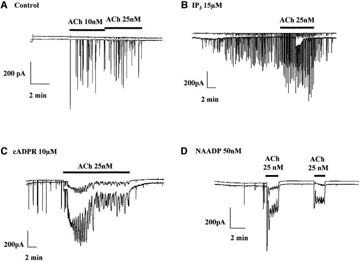
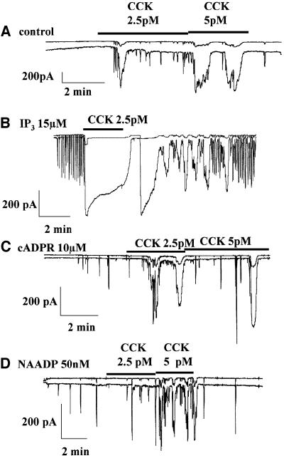
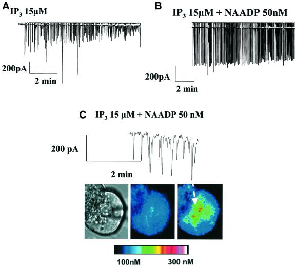
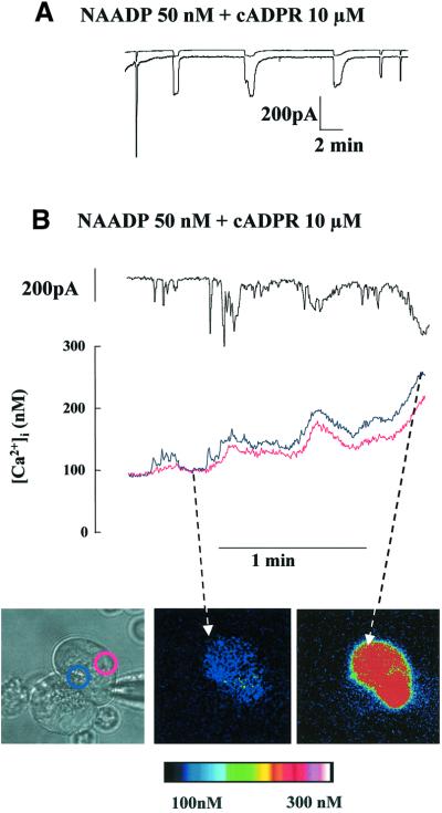
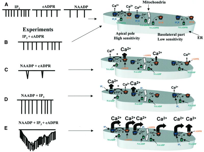
In pancreatic acinar cells, low, threshold concentrations of acetylcholine (ACh) or cholecystokinin (CCK) induce repetitive local cytosolic Ca2+ spikes in the apical pole, while higher concentrations elicit global signals. We have investigated the process that transforms local Ca2+ spikes to global Ca2+ transients, focusing on the interactions of multiple intracellular messengers. ACh-elicited local Ca2+ spikes were transformed into a global sustained Ca2+ response by cyclic ADP-ribose (cADPR) or nicotinic acid adenine dinucleotide phosphate (NAADP), whereas inositol 1,4,5-trisphosphate (IP3) had a much weaker effect. In contrast, the response elicited by a low CCK concentration was strongly potentiated by IP3, whereas cADPR and NAADP had little effect. Experiments with messenger mixtures revealed a local interaction between IP3 and NAADP and a stronger global potentiating interaction between cADPR and NAADP. NAADP strongly amplified the local Ca2+ release evoked by a cADPR/IP3 mixture eliciting a vigorous global Ca2+ response. Different combinations of Ca2+ releasing messengers can shape the spatio-temporal patterns of cytosolic Ca2+ signals. NAADP and cADPR are emerging as key messengers in the globalization of Ca2+ signals.
Figures


Similar articles
-
Two different but converging messenger pathways to intracellular Ca(2+) release: the roles of nicotinic acid adenine dinucleotide phosphate, cyclic ADP-ribose and inositol trisphosphate.EMBO J. 2000 Jun 1;19(11):2549-57. doi: 10.1093/emboj/19.11.2549. EMBO J. 2000. PMID: 10835353 Free PMC article.
-
Specific Ca2+ signaling evoked by cholecystokinin and acetylcholine: the roles of NAADP, cADPR, and IP3.Annu Rev Physiol. 2001;63:99-117. doi: 10.1146/annurev.physiol.63.1.99. Annu Rev Physiol. 2001. PMID: 11181950 Review.
-
Coordination of agonist-induced Ca2+-signalling patterns by NAADP in pancreatic acinar cells.Nature. 1999 Mar 4;398(6722):74-6. doi: 10.1038/18032. Nature. 1999. PMID: 10078532
-
NAADP induces Ca2+ oscillations via a two-pool mechanism by priming IP3- and cADPR-sensitive Ca2+ stores.EMBO J. 2001 Jun 1;20(11):2666-71. doi: 10.1093/emboj/20.11.2666. EMBO J. 2001. PMID: 11387201 Free PMC article.
-
Adenine nucleotide diphosphates: emerging second messengers acting via intracellular Ca2+ release.Am J Physiol. 1996 Oct;271(4 Pt 1):C1007-24. doi: 10.1152/ajpcell.1996.271.4.C1007. Am J Physiol. 1996. PMID: 8897805 Review.
Cited by
-
Ca2+ signalling and membrane current activated by cADPr in starfish oocytes.Pflugers Arch. 2003 Aug;446(5):541-52. doi: 10.1007/s00424-003-1076-1. Epub 2003 May 16. Pflugers Arch. 2003. PMID: 12756567
-
Preferential Coupling of the NAADP Pathway to Exocytosis in T-Cells.Messenger (Los Angel). 2015 Jun;4(1):53-66. doi: 10.1166/msr.2015.1040. Messenger (Los Angel). 2015. PMID: 27330870 Free PMC article.
-
Ca2+ Signaling in Exocrine Cells.Cold Spring Harb Perspect Biol. 2020 May 1;12(5):a035279. doi: 10.1101/cshperspect.a035279. Cold Spring Harb Perspect Biol. 2020. PMID: 31636079 Free PMC article. Review.
-
Fast Ca(2+)-dependent inactivation of the store-operated Ca2+ current (ISOC) in liver cells: a role for calmodulin.J Physiol. 2004 Jul 1;558(Pt 1):85-97. doi: 10.1113/jphysiol.2004.065870. Epub 2004 Apr 30. J Physiol. 2004. PMID: 15226409 Free PMC article.
-
Caffeine protects against experimental acute pancreatitis by inhibition of inositol 1,4,5-trisphosphate receptor-mediated Ca2+ release.Gut. 2017 Feb;66(2):301-313. doi: 10.1136/gutjnl-2015-309363. Epub 2015 Dec 7. Gut. 2017. PMID: 26642860 Free PMC article.
References
-
- Albrieux M., Lee,H.G. and Villaz,M. (1998) Calcium signaling by cyclic ADP-ribose, NAADP and inositol trisphosphate are involved in distinct functions in ascidian oocytes. J. Biol. Chem., 273, 14566–14574. - PubMed
-
- Berridge M.J., Bootman,M.D. and Lipp,P. (1998) Calcium — a life and death signal. Nature, 395, 645–648. - PubMed
-
- Berridge M.J., Lipp,P. and Bootman,M.D. (2000) The versatility and universality of calcium signaling. Nature Rev. Mol. Cell. Biol., 1, 11–21. - PubMed
Publication types
MeSH terms
Substances
LinkOut - more resources
Full Text Sources
Other Literature Sources
Miscellaneous
