. 2002 Mar 1;21(5):942-53.
doi: 10.1093/emboj/21.5.942.

The Tim9p-Tim10p complex binds to the transmembrane domains of the ADP/ATP carrier

Affiliations

The Tim9p-Tim10p complex binds to the transmembrane domains of the ADP/ATP carrier

Sean P Curran et al. EMBO J. .

Abstract

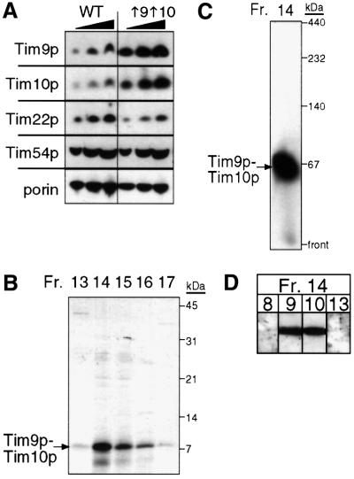
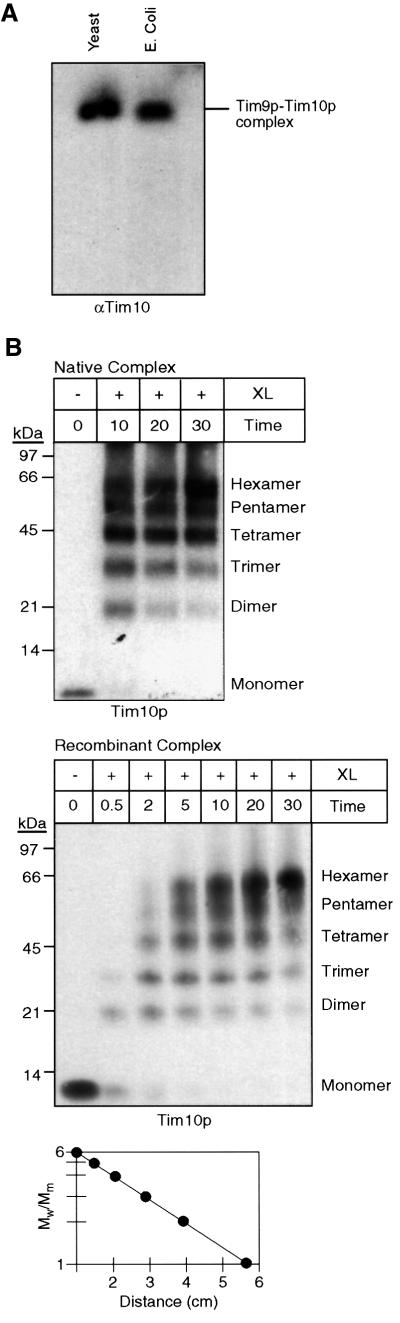
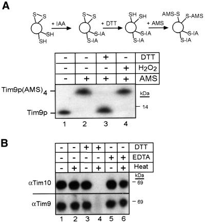
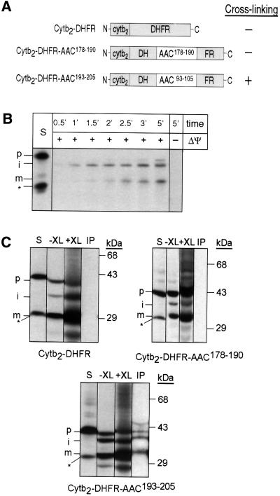
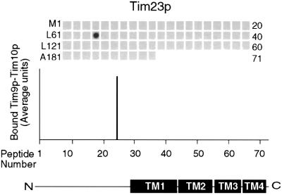
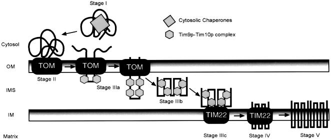
The soluble Tim9p-Tim10p (Tim, translocase of inner membrane) complex of the mitochondrial intermembrane space mediates the import of the carrier proteins and is a component of the TIM22 import system. The mechanism by which the Tim9p-Tim10p complex assembles and binds the carriers is not well understood, but previous studies have proposed that the conserved cysteine residues in the 'twin CX3C' motif coordinate zinc and potentially generate a zinc-finger-like structure that binds to the matrix loops of the carrier proteins. Here we have purified the native and recombinant Tim9p-Tim10p complex, and show that both complexes resemble each other and consist of three Tim9p and three Tim10p. Results from inductively coupled plasma--mass spectrometry studies failed to detect zinc in the Tim9p-Tim10p complex. Instead, the cysteine residues seemingly formed disulfide linkages. The Tim9p-Tim10p complex bound specifically to the transmembrane domains of the ADP/ATP carrier, but had no affinity for Tim23p, an inner membrane protein that is inserted via the TIM22 complex. The chaperone-like Tim9p-Tim10p complex thus may prevent aggregation of the unfolded carrier proteins in the aqueous intermembrane space.


Figures

None
Fig. 1. Purification of the Tim9p–Tim10p complex from a yeast strain overexpressing Tim9p and Tim10p. (A) Tim9p and Tim10p were overexpressed 6- to 8-fold, but the expression of Tim22p and Tim54p was not changed. Mitochondrial proteins (50, 100 and 150 µg) were separated by SDS–PAGE, followed by immunoblot analysis from the parental strain (WT) and a strain (↑9↑10) co-transformed with two 2µ plasmids expressing TIM9 and TIM10, respectively, and analyzed by immunoblotting with monospecific antisera for Tim9p, Tim10p, Tim22p, Tim54p and porin. Proteins were identified by incubation with [125I]protein A; the amount was quantitated by scanning laser densitometry. (B) The Tim9p–Tim10p complex was purified from the mitochondrial intermembrane space. After successive chromatography steps (see Materials and methods), fractions from the final purification step (gel filtration) were analyzed for Tim9p and Tim10p by SDS–PAGE and Coomassie Blue staining. (C) As in (B), fraction 14 was analyzed on a 6–16% blue native gel followed by immunoblotting with antiserum for Tim10p. (D) As in (B), fraction 14 was separated by SDS–PAGE, followed by immunoblot analysis with monospecific antisera for Tim8p, Tim9p, Tim10p and Tim13p. Proteins were identified by incubation with [125I]protein A.
None
Fig. 2. The recombinant and native Tim9p–Tim10p complexes have identical properties. (A) An expression plasmid was constructed in which TIM9 and TIM10 were each placed behind a ribosomal binding site. After transformation into the E.coli host BL21(DE3) and induction, the Tim9p–Tim10p complex (E.coli) was purified (see Materials and methods) and separated on a 6.0% native gel. A mitochondrial intermembrane space fraction (yeast) was also separated. Immunoblot analysis was performed with monospecific antisera against Tim10p (αTim10) and Tim9p (unpublished data). Proteins were identified by incubation with [125I]protein A. (B) For intermolecular cross-linking assays, an intermembrane space fraction (native complex, top blot) and the recombinant Tim9p–Tim10p complex (recombinant complex, bottom blot) were incubated with 0.1% glutaraldehyde (XL) and aliquots were removed at the indicated time points (min). Cross-linked products were separated on 16% tricine gels, followed by immunoblot analysis with monospecific antisera against Tim10p. The number of subunits is indicated on the right. Electrophoretic mobility (graph) of the products from the 10 min time point of cross-linking was plotted against the logarithm of Mw/Mm, where Mw is the relative molecular weight of the cross-linked species and Mm is the molecular mass of the Tim10p or Tim9p monomer (10 kDa).
None
Fig. 3. The sulfhydryl groups on the cysteine residues of the ‘twin CX3C’ motif are occupied. (A) To elucidate the state of the cysteine residues in the Tim9p–Tim10p complex, a thiol-trapping method (Jakob et al., 1999) was used (schematic). A mitochondrial intermembrane space fraction (200 µg per lane) was either left untreated (lane 2), reduced with 10 mM DTT (lane 3) or oxidized with 5% H2O2 (lane 4). The samples were then alkylated with IAA to block free sulfhydryl groups, following treatment with DTT to reduce any disulfide bonds. The remaining free sulfhydryl groups were alkylated with AMS, which creates an increase in molecular mass of 0.5 kDa. The samples were separated by SDS–PAGE, followed by immunoblot analysis with antiserum against Tim10p. As a control, an untreated sample is included (lane 1). Tim9p and Tim9p(AMS)4 are denoted. (B) The reductant DTT interferes with refolding of the Tim9p–Tim10p complex. The recombinant Tim9–Tim10p complex was heated to 95°C in the presence of DTT (lane 4) or EDTA (lane 6) and quickly cooled in an ice bath, followed by blue native gel electrophoresis. As a control, samples were untreated (lane 1), heated (lane 2) or treated with DTT (lane 3) or EDTA (lane 5) and incubated on ice. Immunoblot analysis was performed with an antibody against Tim10p and Tim9p.
None
Fig. 4. Native and recombinant Tim9p–Tim10p complex show a similar binding pattern to a peptide scan derived from the sequence of AAC. (A) The native Tim9p–Tim10p complex (200 nM) purified from the mitochondrial intermembrane space was incubated with a peptide scan consisting of 13mers derived from AAC. The first peptide comprises amino acids 1–13 of the AAC, the second peptide residues 4–16 and the third peptide residues 7–19, etc. The labeling on the left indicates the first amino acid of the left-most peptide of each row. The labeling on the right side indicates the number of the right-most peptide of each row. After washing the membrane, the bound Tim9p–Tim10p complex was transferred to a PVDF membrane, followed by immunoblot analysis with antiserum specific for Tim10p and [125I]protein A. Binding was quantified by scanning laser densitometry from at least three independent experiments and plotted for each peptide. Transmembrane domains and repeat modules of AAC are plotted corresponding to the respective peptides. (B) As in (A), except that 200 nM recombinant Tim9p–Tim10p complex was incubated with the peptide scan.
None
Fig. 5. Tim9p and Tim10p are cross-linked to an AAC peptide placed within a synthetic precursor in the mitochondrial intermembrane space. (A) Diagram of Cytb2–DHFR constructs in which peptides derived from the AAC sequence have been inserted into DHFR. Cytb2–DHFR is a fusion between residues 1–167 of Cytb2 and DHFR. Cytb2–DFHR–AAC178–190 contains AAC residues 178–190 (peptide 60 from Figure 4, which showed no binding). Cytb2–DHFR–AAC193–205 contains AAC residues 193–205 (peptide 65 from Figure 4, which showed binding). (B) The radiolabeled Cytb2–DHFR precursor was synthesized in vitro and incubated for the indicated times at 25°C in the presence and absence of a membrane potential (ΔΨ) with wild-type mitochondria. Samples were treated with protease to remove non-imported precursor and analyzed by SDS–PAGE and fluorography. STD, 25% of the radioactive precursor added to the assay. p, precursor; i, intermediate; m, mature. The asterisk denotes a translation product created by translation initiation from an internal methionine; this product is not imported. (C) The radiolabeled precursor was imported into wild-type mitochondria for 10 min at 25°C (–XL). The chemical cross-linker DSS (+XL) was added at 1 mM. After quenching, the sample was denatured and immunoprecipitated with antibodies against Tim9p and Tim10p (IP). Bound proteins were eluted with SDS-containing sample buffer and analyzed by SDS–PAGE and fluorography. S, 10% of the amount of precursor that was added to the non-cross-linked import assay. p, precursor; i, intermediate; m, mature. The asterisk denotes a translation product created by translation initiation from an internal methionine; this product is not imported.
None
Fig. 6. Native Tim9p–Tim10p complex does not bind to Tim23p. The native Tim9p–Tim10p complex (200 nM) purified from the mitochondrial intermembrane space was incubated with a peptide scan consisting of 13mers derived from Tim23p, as in Figure 4. Binding was quantified by scanning laser densitometry from at least three independent experiments and plotted for each peptide. Transmembrane domains of Tim23p are plotted corresponding to the respective peptides.
None
Fig. 7. Proposed model for the role of the Tim9p–Tim10p complex in the import of carrier proteins. Carrier proteins are escorted by cytosolic chaperones (stage I) to receptors of the TOM complex (stage II). The precursor passes through the TOM channel to the intermembrane space and the Tim9p–Tim10p complex binds to hydrophobic residues in the transmembrane domains of the carriers (stage IIIa). In the aqueous intermembrane space, the Tim9p–Tim10p complex maintains the carrier in an import-competent state (stage IIIb) and delivers it to the TIM22 translocon (stage IIIc). The TIM22 translocon mediates insertion into the inner membrane (stage IV) and then the carrier assembles into a functional dimer (stage V).

References

    1. Adam A., Endres,M., Sirrenberg,C., Lottspeich,F., Neupert,W. and Brunner,M. (1999) Tim9, a new component of the TIM22.54 translocase in mitochondria. EMBO J., 18, 313–319. - PMC - PubMed
    1. Azem A., Weiss,C. and Goloubinoff,P. (1998) Structural analysis of GroE chaperonin complexes using chemical cross-linking. Methods Enzymol., 290, 253–268. - PubMed
    1. Bauer M.F., Hofmann,S., Neupert,W. and Brunner,M. (2000) Protein translocation into mitochondria: the role of TIM complexes. Trends Cell Biol., 10, 25–31. - PubMed
    1. Brix J., Rudiger,S., Bukau,B., Schneider-Mergener,J. and Pfanner,N. (1999) Distribution of binding sequences for the mitochondrial import receptors Tom20, Tom22 and Tom70 in a presequence-carrying preprotein and a non-cleavable preprotein. J. Biol. Chem., 274, 16522–16530. - PubMed
    1. Brix J., Ziegler,G.A., Dietmeier,K., Schneider-Mergener,J., Schulz,G.E. and Pfanner,N. (2000) The mitochondrial import receptor Tom70: identification of a 25 kDa core domain with a specific binding site for preproteins. J. Mol. Biol., 303, 479–488. - PubMed

Publication types

MeSH terms

Substances