. 2002 Mar 1;21(5):966-75.
doi: 10.1093/emboj/21.5.966.

The Wnt antagonist Dickkopf-1 is regulated by Bmp signaling and c-Jun and modulates programmed cell death

Affiliations

The Wnt antagonist Dickkopf-1 is regulated by Bmp signaling and c-Jun and modulates programmed cell death

Lars Grotewold et al. EMBO J. .

Abstract

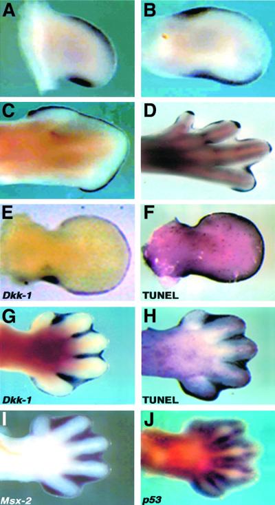
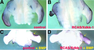
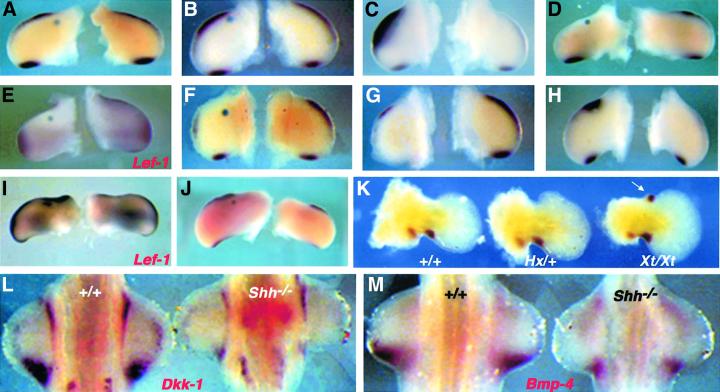
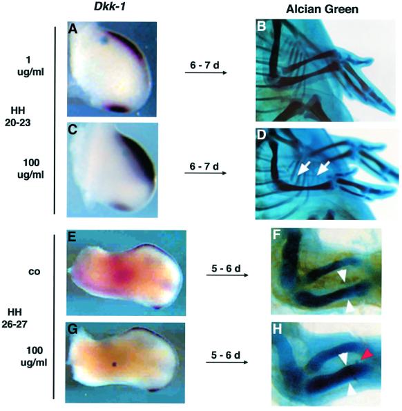
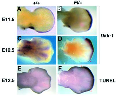
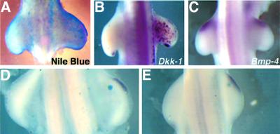
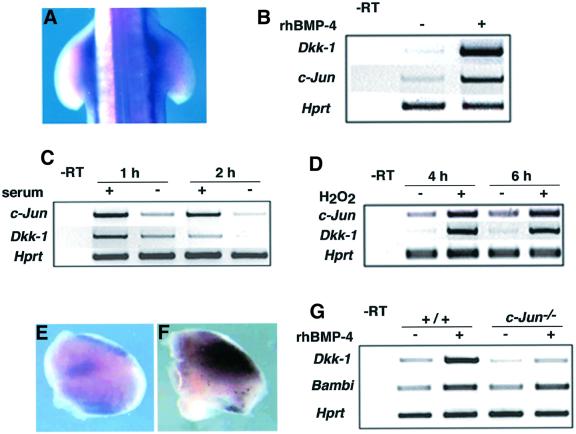
Dickkopf-1 (Dkk-1) has been shown to be a potent inhibitor of Wnt/beta-catenin signaling in a variety of assays and organisms. In this study, we show that expression of Dkk-1 overlaps significantly with the sites of programmed cell death in normal as well as mutant vertebrate limb development, and identify several of its upstream regulators, one of which is Bmp-4. Interestingly, Bmp-4 only activates Dkk-1 when it concomitantly induces apoptosis. Moreover, Dkk-1 is heavily up-regulated by UV irradiation and several other genotoxic stimuli. We further show that normal expression of Dkk-1 is dependent on the Ap-1 family member c-Jun and that overexpression of Dkk-1 enhances Bmp-triggered apoptosis in the vertebrate limb. Taken together, our results provide evidence for an important role of Dkk-1-mediated inhibition of Wnt/beta-catenin signaling in response to different stress signals that all converge on the activation of c-Jun in vivo.


Figures

None
Fig. 1. Dkk-1 is expressed at sites of apoptosis during vertebrate limb development. (A–D) Chick limb buds of different stages, (A) HH23 wing, (B) HH25 wing, (C) HH32 wing and (D) HH32 leg, show Dkk-1 transcripts in the ANZ, PNZ, the AER and INZ and the developing joints. (EDkk-1 expression in an E11.5 mouse forelimb bud in the AER and posterior mesenchyme and (F) whole-mount TUNEL staining of an age-matched forelimb bud indicates the co-localization of Dkk-1 transcripts and the sites of apoptosis. (G) At E13.5, Dkk-1 expression is confined to the interdigital mesenchyme, where massive apoptosis takes place as the TUNEL staining of an E13.5 limb bud indicates (H). At this time point Dkk-1 is co-expressed with the pro-apoptotic genes Msx-2 (I) and p53 (J).
None
Fig. 2. Dkk-1 promotes apoptosis in limb buds. (A and B) TUNEL stainings of RCAS-infected wing buds, ventral view. (A) Control wing infected with RCAS/AP showing the normal pattern of PCD in the ANZ and AER. (B) Dramatic truncation of a wing bud infected with RCAS/Xdkk-1. Note the massive apoptosis in the truncated wing compared with the contralateral control wing. (C and D) Dorsal views of wing buds infected with RCAS/AP (C) and RCAS/Xdkk-1 (D), which additionally received a bead soaked in rhBMP-4 (arrows). (C) Twenty hours after bead implantation the ectopic Bmp signal leads to an increased PCD in a region surrounding the bead as shown by TUNEL. (D) The overexpression of Dkk-1 dramatically enhances this effect of Bmp. The region of cells undergoing PCD covers nearly the complete wing mesoderm.
None
Fig. 3. Regulation of Dkk-1 by Bmp, Fgf and Shh. Surgical manipulations of wing buds in (A)–(K) were performed between HH20 and HH23. Operated wings (left) and contralateral controls (right) are presented in each panel. (A) A BSA-soaked control bead or (B) a bead soaked in 1 µg/ml rhBMP-4 does not affect Dkk-1 expression after 8 h. (C) Massive ectopic expression of Dkk-1 around a Bmp bead (100 µg/ml) 2 h post-implantation. Note that Dkk-1 is only induced between the bead and the AER. (D) Twenty hours after application of 100 µg/ml rhBMP-4, Dkk-1 expression is completely down-regulated. (ELef-1 expression is drastically reduced 8 h after the same treatment. (F) Loss of Dkk-1 transcripts in the vicinity of a bead soaked in 1 mg/ml Noggin after 8 h. (G) Removal of the AER leads to a decline of Dkk-1 transcripts in the subjacent mesenchyme after 6 h. (HDkk-1 is induced by 1 mg/ml Fgf-8 (8 h). (ILef-1 expression is also clearly enhanced following this treatment. (J) Shh (2.5 mg/ml) has no effect on Dkk-1 expression, even after 24 h. Note the increased size of the operated wing, which indicates that the Shh protein provided by the bead is functional. (KDkk-1 expression in E12.0 mouse forelimb buds. Wild type (left) and Hx/+ (middle) show the same pattern, whereas a strong ectopic expression domain of Dkk-1 is detectable in the anterior mesenchyme of Xt/Xt (right) limb buds (arrow). (LDkk-1 expression is drastically reduced in Shh–/– forelimb buds (right) at E10.5 (left side shows an age-matched wild-type control). (MBmp-4 is also down-regulated in Shh–/– limb buds (right) compared with wild type (left) at E10.5.
None
Fig. 4. Bmp-4 concomitantly induces Dkk-1 expression and apoptosis. The bead implantations in (A)–(D) have been carried out between HH20 and HH23, and those in (E)–(H) at HH26/27. (A) A control bead soaked in 1 µg/ml rhBMP-4 does not affect Dkk-1 expression. (B) Wild-type skeletal pattern of a wing treated the manner described in (A) after 10 days of incubation. (C) rhBMP-4 100 µg/ml rapidly induces Dkk-1 expression, resulting in the complete absence of the radius after 10 days [(D), arrows)]. (E) Control (co) wing shows the wild-type pattern of Dkk-1 expression at HH26. (F) Wild-type size of radius and ulna in an E10 embryo. (G) A Bmp bead (100 µg/ml) implanted into the center of a HH26/27 wing does not induce Dkk-1 expression after 8 h. (H) This treatment leads to a significant increase in bone mass of radius and ulna after 10 days. White arrowheads have the same distance as in (F), the red arrowhead points to the implanted bead.
None
Fig. 5. Dkk-1 expression and PCD in Ft/+ forelimb buds. (A) E11.5 wild-type limb bud shows Dkk-1 transcripts in the PNZ and AER. (B) Ectopic expression of Dkk-1 in the distal-most mesenchyme of an E11.5 Ft/+ limb bud. (CDkk-1 expression in the interdigital mesenchyme of an E12.5 wild-type limb bud. (DDkk-1 is ectopically activated at E12.5 in Ft/+ forelimb buds (red arrowheads). (E) TUNEL staining shows the wild-type pattern of PCD at E12.5. Apoptotic cells are restricted to the AER and the distal-most interdigital mesenchyme. (F) High ectopic cell death in Ft/+ forelimb bud (red arrowheads) in a region that significantly overlaps with that of ectopic Dkk-1 transcription [compare with (D)].
None
Fig. 6. Dkk-1 is induced by UV irradiation and staurosporin. (A) Nile Blue staining of a chick embryo 10 h after UV irradiation. Note the massive cell death in the right irradiated limb bud. The non-irradiated left side served as an internal control in all of these experiments. (BDkk-1 is ectopically expressed in a salt-and-pepper-like fashion 8 h after UV irradiation. (CBmp-4 expression is not altered after 8 h. (D and E) Beads soaked in staurosporin induce Dkk-1 expression after 2 h (D) and 4 h (E).
None
Fig. 7. Dkk-1 is regulated by c-Jun. (A) c-Jun is up-regulated 1 h after UV irradiation. Note the intense c-Jun expression in the right wing bud and the flank compared with the non-irradiated left side of the embryo. (BDkk-1 and c-Jun are co-induced by application of rhBMP-4 to MEF after 30 min. Hprt was used for standardization in all RT–PCR experiments. (C) c-Jun and Dkk-1 are down-regulated in MEF, 1 and 2 h after serum withdrawal. (D) H2O2 strongly up-regulates c-Jun and Dkk-1. (E and F) Limb bud explants electroporated with a control vector (E) and a c-Jun expression plasmid (F). Note the intense up-regulation of Dkk-1 expression 6 h after electroporation of the c-Jun expression vector (F). (GDkk-1 expression is reduced by 40% in c-Jun–/– MEF compared with wild type (compare +/+ and c-Jun–/– without rhBMP-4). After the application of rhBMP-4 Dkk-1 is 5.2-fold induced in wild-type MEF, but only 1.7-fold in c-Jun–/– cells. Bambi is normally expressed and induced by the same factor in c-Jun–/– as in wild-type MEF. –RT, control without reverse transcriptase.

Similar articles

Cited by

References

    1. Adachi-Yamada T., Fujimura-Kamada,K., Nishida,Y. and Matsumoto,K. (1999) Distortion of proximodistal information causes JNK-dependent apoptosis in Drosophila wing. Nature, 400, 166–169. - PubMed
    1. Angel P., Hattori,K., Smeal,T. and Karin,M. (1988) The jun proto-oncogene is positively autoregulated by its product, Jun/AP1. Cell, 55, 875–885. - PubMed
    1. Behrens A., Sibilia,M. and Wagner,E.F. (1999) Amino-terminal phosphorylation of c-Jun regulates stress-induced apoptosis and cellular proliferation. Nature Genet., 21, 326–329. - PubMed
    1. Bejsovec A. (2000) Wnt signaling: an embarrassment of receptors. Curr. Biol., 10, R919–R922. - PubMed
    1. Bossy-Wetzel E., Bakiri,L. and Yaniv,M. (1997) Induction of apoptosis by the transcription factor c-Jun. EMBO J., 16, 1695–1709. - PMC - PubMed

Publication types

MeSH terms

Substances