Comparative Study
. 2002 Mar 1;21(5):1012-20.
doi: 10.1093/emboj/21.5.1012.

The C. elegans ric-3 gene is required for maturation of nicotinic acetylcholine receptors

Affiliations
Comparative Study

The C. elegans ric-3 gene is required for maturation of nicotinic acetylcholine receptors

Sarah Halevi et al. EMBO J. .

Abstract

Mutations in ric-3 (resistant to inhibitors of cholinesterase) suppress the neuronal degenerations caused by a gain of function mutation in the Caenorhabditis elegans DEG-3 acetylcholine receptor. RIC-3 is a novel protein with two transmembrane domains and extensive coiled-coil domains. It is expressed in both muscles and neurons, and the protein is concentrated within the cell bodies. We demonstrate that RIC-3 is required for the function of at least four nicotinic acetylcholine receptors. However, GABA and glutamate receptors expressed in the same cells are unaffected. In ric-3 mutants, the DEG-3 receptor accumulates in the cell body instead of in the cell processes. Moreover, co-expression of ric-3 in Xenopus laevis oocytes enhances the activity of the C.elegans DEG-3/DES-2 and of the rat alpha-7 acetylcholine receptors. Together, these data suggest that RIC-3 is specifically required for the maturation of acetylcholine receptors.


Figures

None
Fig. 1. Cloning and molecular analysis of ric-3. (A) Genetic map of chromosome IV showing the position of ric-3 relative to nearby genes. (B) Physical map of cosmid T14A8, showing positions of predicted genes (T14A8.1, which encodes ric-3, is in bold) and of restriction sites used in this study. (C) Structure of the ric-3 gene. Exon structure was determined from the analysis of four cDNAs. Blocks, exons; bold line, non-translated regions; thin line, 5′ upstream non-transcribed region. The alternatively spliced exon is marked with an asterisk. Sites of mutation in ric-3 are indicated: md1013, md1034, md1181, md1272, md1274, md1286 and md1442 are Tc1 insertions, hm9, hm19, hm54 and hm65 are nonsense mutations, and md146, md156, md158 and md226 are deletions (12.5 kb, 7 bp, 5 bp and 12 bp, respectively). (D) Sequence of the RIC-3 protein derived from cDNAs (yk704a8 and yk722a6). Single underlines indicate transmembrane regions, double underlines indicate coiled-coil regions, and the dashed line indicates amino acids that are absent in the alternatively spliced cDNA variants (yk719f3 and yk266d12), in which the last G in this segment is converted to an R.
None
Fig. 2. A RIC-3 homolog. (A) Homology with Drosophila CG9349 protein in the region spanning the transmembrane domains. Underlines indicate putative transmembrane domains. (B) Structure and topology predictions for RIC-3 and CG9349. Barrels indicate positions of coiled-coil regions. CG9349 has only one predicted coiled-coil domain.
None
Fig. 3. Localization of RIC-3. RIC-3 localization was visualized using a RIC-3::GFP functional fusion. (A) Head region, showing the pharyngeal muscles (the double-lobed organ in the middle) and head ganglia neurons seen between the two lobes of the pharynx. Confocal section, scale bar: 10 µM. (B) Body muscles and neurons. The row of neurons in the middle marked by asterisks are motor neurons, on both sides of which are seen the body muscles. In the left corner, marked by an arrow, are the posterior–lateral ganglion neurons showing strong fluorescence in cell bodies and weak fluorescence of processes. Confocal section, scale bar: 50 µM. (C) RIC-3 localization in the body muscles as visualized using a myo-3ric-3gfp fusion. Scale bar: 20 µM.
None
Fig. 4. Defective cholinergic transmission in the C.elegans pharyngeal muscle. Representative traces for the electrical activity of the pharynx of wild-type (N2) and ric-3(hm9) mutant animals. MC EPSP is the excitatory cholinergic activity, seen as a small spike preceding muscle depolarization (large upward spike). M3 IPSP is the inhibitory glutamatergic activity, seen as a series of small spikes preceding muscle repolarization (large downward spike). In the ric-3(hm9) mutant, the small depolarizatory current seen in the beginning of the trace may represent I phase activity, i.e. a sub-threshold MC spike.
None
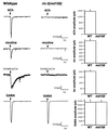
Fig. 5. Defective response to acetylcholine receptor agonists in the body muscles. Electrical responses of wild-type and mutant ric-3(md158) body muscles to puffs of ACh, nicotine, levamisole or GABA (100 µM each). On the left are representative traces of the responses; arrows indicate the time of agonist application. On the right is shown the average of the responses.
None
Fig. 6. DEG-3 accumulates in cell bodies of ric-3 mutants. Immunohistochemical analysis using DEG-3 antibodies on young adults. Shown is the PVD neuron and processes in (A) wild type and (Bric-3(md158). Note the size difference between wild type and ric-3, both of which are young adults. Confocal section, scale bar: 20 µM.
None
Fig. 7. RIC-3 co-expression enhances acetylcholine receptor activity in Xenopus oocytes. Amplitude of choline- (3.2 mM) or glutamate- (1 mM) dependent currents measured in oocytes together with (striped) or without (filled) co-expression of RIC-3. The DEG-3/DES-2 experiments represent n = 38 (oocytes), N = 6 (frogs) each. α-7, n = 23, N = 4; GluR3, n = 7, N = 2; and RIC-3 alone, n = 10, N = 3. The effects of co-expressing RIC-3 on DEG-3/DES-2- and α-7-dependent current amplitudes are significant using a paired t-test at >99%.

References

    1. Altschul S.F., Gish,W., Miller,W., Myers,E.W. and Lipman,D.J. (1990) Basic local alignment search tool. J. Mol. Biol., 215, 403–410. - PubMed
    1. Baker E.K., Colley,N.J. and Zuker,C.S. (1994) The cyclophilin homolog ninaA functions as a chaperone, forming a stable complex in vivo with its protein target rhodopsin. EMBO J., 13, 4886–4895. - PMC - PubMed
    1. Bargmann C.I. (1998) Neurobiology of the Caenorhabditis elegans genome. Science, 282, 2028–2033. - PubMed
    1. Berger B., Wilson,D.B., Wolf,E., Tonchev,T., Milla,M. and Kimm,P.S. (1995) Predicting coiled coils by use of pairwise residue correlations. Proc. Natl Acad. Sci. USA, 92, 8259–8263. - PMC - PubMed
    1. Blount P. and Merlie,J.P. (1991) BIP associates with newly synthesized subunits of the mouse muscle nicotinic receptor. J. Cell Biol., 113, 1125–1132. - PMC - PubMed

Publication types

MeSH terms