. 2002 Mar 1;21(5):1031-40.
doi: 10.1093/emboj/21.5.1031.

Conversion of raft associated prion protein to the protease-resistant state requires insertion of PrP-res (PrP(Sc)) into contiguous membranes

Affiliations

Conversion of raft associated prion protein to the protease-resistant state requires insertion of PrP-res (PrP(Sc)) into contiguous membranes

Gerald S Baron et al. EMBO J. .

Abstract

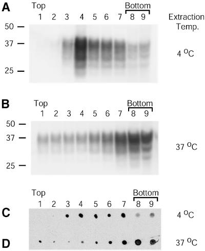
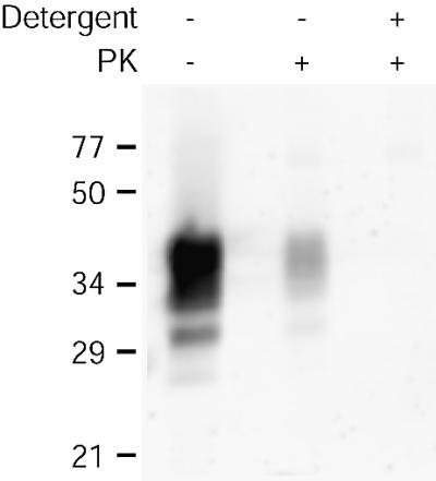
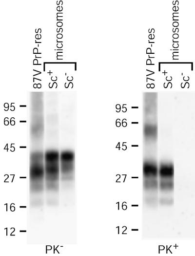
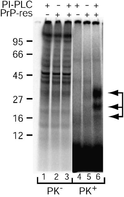
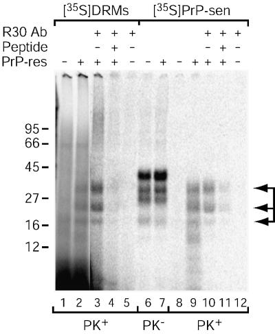
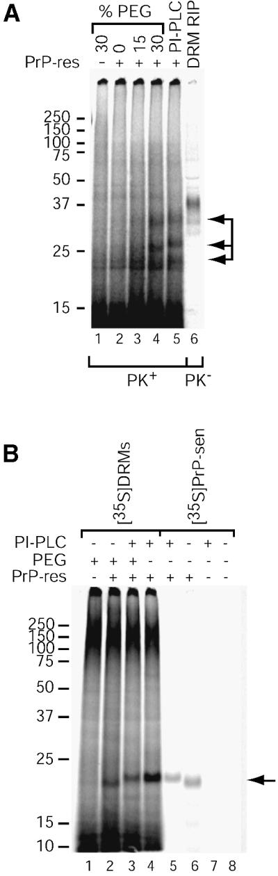
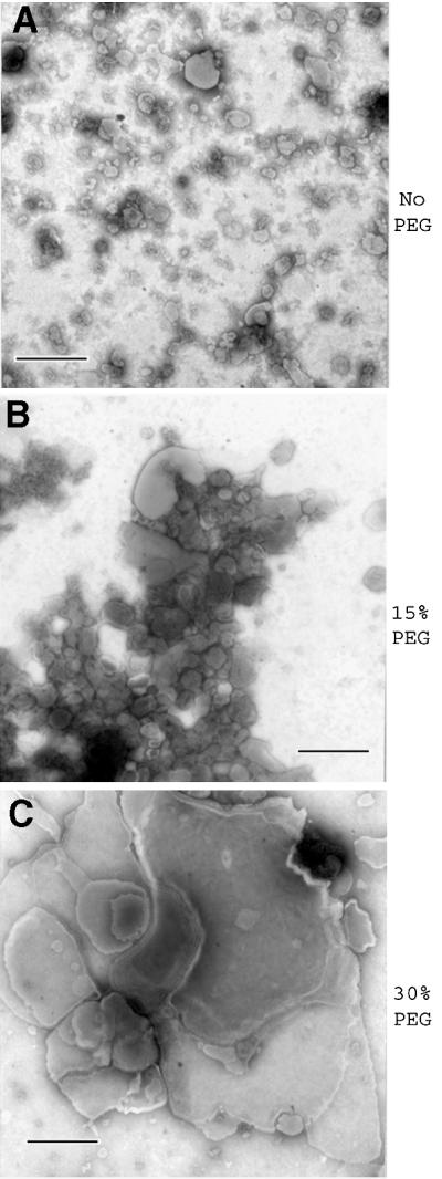
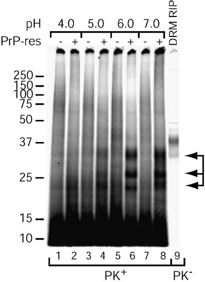
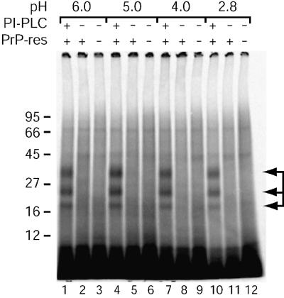
Prion protein (PrP) is usually attached to membranes by a glycosylphosphatidylinositol-anchor that associates with detergent-resistant membranes (DRMs), or rafts. To model the molecular processes that might occur during the initial infection of cells with exogenous transmissible spongiform encephalopathy (TSE) agents, we examined the effect of membrane association on the conversion of the normal protease-sensitive PrP isoform (PrP-sen) to the protease-resistant isoform (PrP-res). A cell-free conversion reaction approximating physiological conditions was used, which contained purified DRMs as a source of PrP-sen and brain microsomes from scrapie-infected mice as a source of PrP-res. Interestingly, DRM-associated PrP-sen was not converted to PrP-res until the PrP-sen was either released from DRMs by treatment with phosphatidylinositol-specific phospholipase C (PI-PLC), or the combined membrane fractions were treated with the membrane-fusing agent polyethylene glycol (PEG). PEG-assisted conversion was optimal at pH 6--7, and acid pre-treating the DRMs was not sufficient to permit conversion without PI-PLC or PEG, arguing against late endosomes/lysosomes as primary compartments for PrP conversion. These observations raise the possibility that generation of new PrP-res during TSE infection requires (i) removal of PrP-sen from target cells; (ii) an exchange of membranes between cells; or (iii) insertion of incoming PrP-res into the raft domains of recipient cells.


Figures

None
Fig. 1. PrP-sen is enriched in DRMs. Mouse neuroblastoma cells (5E4E) overexpressing wild-type mouse PrP were lysed in CBS (pH 6.0) with 1% Triton X-100 at either 4 or 37°C. Lysates were subjected to floatation on Optiprep gradients. Gradient fractions were collected and examined by immunoblotting for the presence of PrP (A and B) and the DRM marker ganglioside GM1 (C and D). Fraction numbers are indicated above each lane. Molecular mass markers are indicated in kDa on the left. The data are representative of two independent experiments.
None
Fig. 2. PrP-sen is surface localized in purified DRMs. DRMs were digested with PK in the presence or absence of detergent (1% Triton X-100) at 37°C. Samples were assayed for PrP by immunoblotting. Results are representative of two independent experiments, each performed in duplicate.
None
Fig. 3. Immunoblot analysis of PrP in mouse brain microsomes. Crude microsomes were prepared from the brains of normal (Sc) or 87V scrapie-infected (Sc+) mice and assayed for total PrP (PK) or PrP-res (PK+) by immunoblotting of untreated or PK-digested samples. Samples of PrP-res purified from 87V-infected mice (87V PrP-res) without (PK) and with (PK+) PK digestion are shown for comparison.
None
Fig. 4. PI-PLC assists cell-free conversion of DRM-associated PrP-sen. Cell-free conversion reactions were performed using [35S]DRMs and crude microsomes from the brains of normal (PrP-res negative lanes) or scrapie-infected (PrP-res positive lanes) mice. PI-PLC was added to the reactions where indicated. PK lanes contain a one-tenth aliquot of each reaction mixture before PK digestion and are shown at reduced contrast to allow visualization of individual bands. PK+ lanes contain a nine-tenths aliquot of each reaction mixture after PK digestion. Arrows indicate PK-resistant [35S]PrP-res bands (lane 6). The data are representative of several independent experiments.
None
Fig. 5. The [35S]DRM conversion product is PrP. Conversion reactions were performed as in Figure 4 using microsomes and [35S]DRMs (with PI-PLC; lanes 1–5) or PI-PLC-digested [35S]PrP-sen (lanes 6–12). Conversion reaction products were immunoprecipitated using either R30 antiserum against a mouse PrP synthetic peptide (89–103; lanes 3, 5, 10 and 12) or R30 pre-absorbed with peptide 89–103 (lanes 4 and 11). Conversion reactions without immunoprecipitation (lanes 1, 2 and 6–9) are shown for comparison. PK and PK+ indicate lanes without and with PK digestion, respectively. PK+ lanes contain nine times the reaction equivalents loaded in PK lanes, except for the immunoprecipitated samples where the entire reaction was PK digested. Arrows indicate PK-resistant [35S]PrP-res bands (lanes 2, 3, 9 and 10). The data are representative of a single experiment performed in duplicate.
None
Fig. 6. PEG assists cell-free conversion of DRM-associated PrP-sen. (A) PEG-assisted [35S]DRM conversion reactions. Samples were treated with various concentrations of PEG or PI-PLC as indicated and cell-free conversion was performed as in Figure 4. PrP-sen was immunoprecipitated from one-fifth equivalents of the [35S]DRMs added to the reaction for comparison (DRM RIP, lane 6). The data are representative of several independent experiments, each performed in duplicate. Arrows indicate the PK-resistant [35S]PrP-res bands (lanes 4 and 5). (B) Effect of PNGase F and PI-PLC treatment. Cell-free conversions were performed using [35S]DRMs treated with 30% PEG or PI-PLC (lanes 1–4). Where indicated, PEG-treated [35S]DRM reactions were also digested with PI-PLC after conversion to demonstrate the PEG-assisted conversion product is GPI anchored (lane 3). For comparison, conversion reactions using purified 87V PrP-res and purified PI-PLC-digested or GPI-anchored [35S]PrP-sen are shown (lanes 5–8). All reactions were deglycosylated with PNGase F after conversion. An arrow indicates the [35S]PrP-res bands (lanes 2–6).
None
Fig. 7. PEG induces membrane fusion in DRM/microsome mixtures. Mixtures of DRMs and normal brain microsomes were subjected to the designated PEG treatment protocol used in the conversion reactions and analyzed by negative staining EM. Bar = 500 nm.
None
Fig. 8. pH dependence of PEG-assisted [35S]DRM conversion reactions. Conversion reactions with microsomes and [35S]DRMs and 30% PEG treatment were performed as in Figure 6. Following PEG treatment, the reactions were incubated in buffers of decreasing pH and assayed for PrP-res. PrP-sen was immunoprecipitated from one-fifth equivalents of the [35S]DRMs added to the reaction for comparison (DRM RIP, lane 9). Arrows indicate the PK-resistant [35S]PrP-res bands (lanes 4, 6 and 8). The data are representative of two independent experiments, each performed in duplicate.
None
Fig. 9. Acid pre-treatment does not facilitate cell-free conversion of DRM-associated PrP-sen. [35S]DRMs were incubated in buffers of decreasing pH, washed in CBS and used in cell-free conversion reactions as in Figure 4. Arrows indicate the PK-resistant [35S]PrP-res bands (lanes 1, 4, 7 and 10). The data are representative of a single experiment performed in duplicate.
None
Fig. 10. Possible mechanisms for transmission of PrP-res (squares) from an infected cell (thick membrane) to an uninfected cell (thin membrane). (A) Exchange of PrP-res-containing membrane microparticles. (B) GPI-anchor-dependent ‘painting’. PrP-sen (shaded circles).

References

    1. Arnold J.E., Tipler,C., Laszlo,L., Hope,J., Landon,M. and Mayer,R.J. (1995) The abnormal isoform of the prion protein accumulates in late-endosome-like organelles in scrapie-infected mouse brain. J. Pathol., 176, 403–411. - PubMed
    1. Batista F.D., Iber,D. and Neuberger,M.S. (2001) B cells acquire antigen from target cells after synapse formation. Nature, 411, 489–494. - PubMed
    1. Bessen R.A., Raymond,G.J. and Caughey,B. (1997) In situ formation of protease-resistant prion protein in transmissible spongiform encephalopathy-infected brain slices. J. Biol. Chem., 272, 15227–15231. - PubMed
    1. Borchelt D.R., Taraboulos,A. and Prusiner,S.B. (1992) Evidence for synthesis of scrapie prion proteins in the endocytic pathway. J. Biol. Chem., 267, 16188–16199. - PubMed
    1. Brown D.A. and Rose,J.K. (1992) Sorting of GPI-anchored proteins to glycolipid-enriched membrane subdomains during transport to the apical cell surface. Cell, 68, 533–544. - PubMed

Publication types

MeSH terms