Comparative Study
. 2002 Mar 1;21(5):1054-62.
doi: 10.1093/emboj/21.5.1054.

Structure of a functional IGF2R fragment determined from the anomalous scattering of sulfur

Affiliations
Comparative Study

Structure of a functional IGF2R fragment determined from the anomalous scattering of sulfur

James Brown et al. EMBO J. .

Abstract

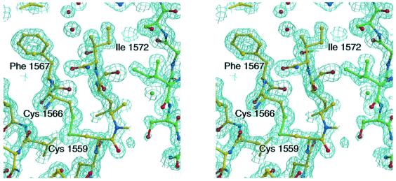
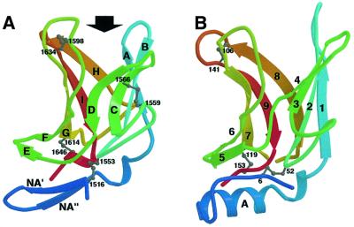
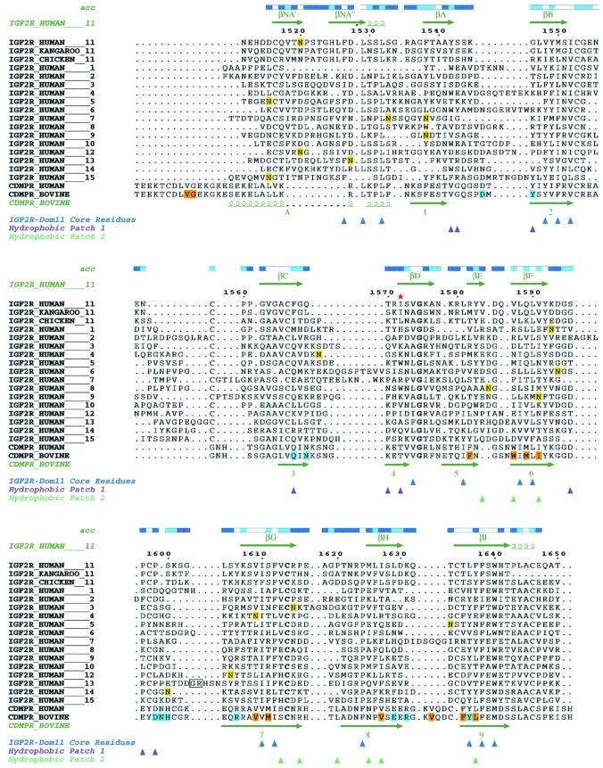
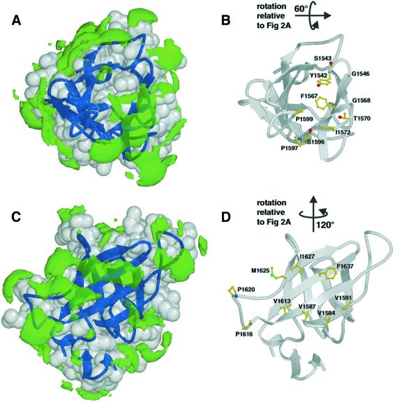
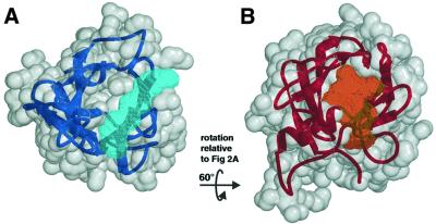
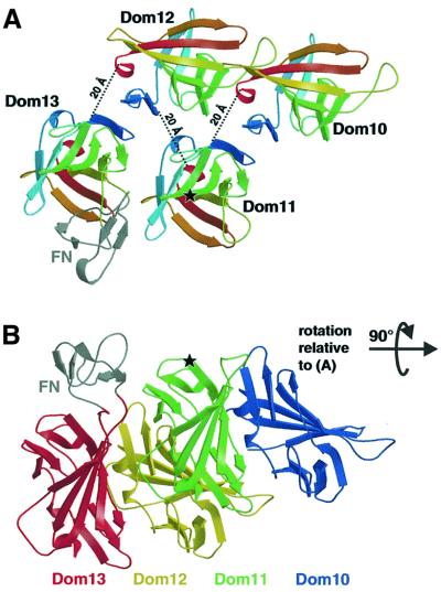
Insulin-like growth factor II receptor (IGF2R) is a multifunctional cell surface receptor implicated in tumour suppression. Its growth inhibitory activity has been associated with an ability to bind IGF-II. IGF2R contains 15 homologous extracellular domains, with domain 11 primarily responsible for IGF-II binding. We report a 1.4 A resolution crystal structure of domain 11, solved using the anomalous scattering signal of sulfur. The structure consists of two crossed beta-sheets forming a flattened beta-barrel. Structural analysis identifies the putative IGF-II binding site at one end of the beta-barrel whilst crystal lattice contacts suggest a model for the full-length IGF2R extracellular region. The structure factors and coordinates of IGF2R domain 11 have been deposited in the Protein Data Bank (accession codes 1GP0 and 1GP3).


Figures

None
Fig. 1. Stereodiagram of the final 2FoFc electron density map for the tetragonal crystal form of IGF2R-Dom11, contoured at 1.7σ. Residues Ile1572 and Phe1567 are in the putative IGF-II binding site (hydrophobic patch 1; see text). A crystal contact molecule is shown as green balls-and-sticks demonstrating the packing of hydrophobic patch 1 against hydrophobic patch 2 (around Ile1627).
None
Fig. 2. Comparison of IGF2R-Dom11 and CDMPR structures. (A) Ribbon diagram of IGF2R-Dom11 showing the flattened β-barrel structure capped off by a β-hairpin. An arrow indicates the putative IGF-II binding region. Ribbons are coloured from blue at the N-terminus to red at the C-terminus and disulfide linkages are represented by grey balls-and-sticks. (B) Ribbon diagram of CDMPR (PDB accession code 1C39) coloured using the same scheme as in (A).
None
Fig. 3. Alignments of human, kangaroo and chicken IGF2R domain 11 sequences, human IGF2R domains 1–10 and 12–15 and human and bovine CDMPR. The IGF2R sequences were aligned using Clustal_W with manual modifications using Jalview, while the human IGF2R-Dom11 and bovine CDMPR were aligned based on structure. Numbering is that of mature human IGF2R-Dom11. Accessibilities of IGF2R-Dom11 residues are given above the alignments (blue, accessible; cyan, intermediate; white, buried). Secondary structure assignments for IGF2R-Dom11 and bovine CDMPR are displayed above and below the alignments, respectively. Potential glycosylation sites in IGF2R are highlighted in yellow, residues involved in the ligand binding pocket of CDMPR are highlighted in cyan and residues involved in CDMPR dimerization are highlighted in orange. In domain 13, the box indicates the residues between which the fibronection-like sequence is inserted. A red star indicates Ile1572 in the IGF-II binding site. Coloured triangles indicate residues of IGF2R-Dom11 that form the core (blue) or that contribute to the hydrophobic surface patches 1 (purple) and 2 (turquoise).
None
Fig. 4. Hydrophobic patches on the surface of IGF2R-Dom11. Views showing (A) hydrophobic patch 1, the putative IGF-II binding region and (B) the residues forming this patch (including Ile1572). Views showing (C) hydrophobic patch 2, an area partially equivalent to the dimerization region of CDMPR, and (D) the residues responsible for this patch. Hydrophobic patches (green) are defined using the program GRID (Goodford, 1985) and are shown here as volumes of pseudo-energies contoured at –2.3 kcal/mol. IGF2R-Dom11 is shown as a grey surface with dark blue secondary structure.
None
Fig. 5. Ligand binding cavities for IGF2R-Dom11 and CDMPR. (A) The proposed IGF-II binding cavity (pale blue) of IGF2R-Dom11 (grey surface and dark blue secondary structure) and (B) the mannose 6-phosphate binding cavity (orange) of CDMPR (grey surface and red secondary structure). Cavities were defined by calculating volumes accessible to the centre of a 1.4 Å radius probe but not to a 6 Å probe, selecting the appropriate cavity and expanding these probe centres by spheres of 1.4 Å radius using VOLUMES (R.M.Esnouf, unpublished program).
None
Fig. 6. Tentative model for the relative arrangement of IGF2R domains 10–13. (A) View looking down on to the putative IGF-II binding site around residue Ile1572 (indicated by a star), showing each domain coloured from blue at the N-terminus to red at the C-terminus. The fibronectin-like insert in domain 13 (coloured grey and labelled FN) was obtained from the Protein Data Bank (accession code 1J7M), edited and positioned approximately at the insertion site using O (Jones et al., 1991). Dotted lines give the distances between the final cysteine of one domain and the first cysteine of the subsequent domain. (B) View at 90° from that in (A) showing the proximity between our proposed IGF-II binding site (again indicated by a star) and the fibronectin-like insert (coloured grey and labelled FN). Domains are coloured from blue (domain 10) to red (domain 13).

Similar articles

Cited by

References

    1. Brünger A.T. et al. (1998) Crystallography and NMR system: A new software suite for macromolecular structure determination. Acta Crystallogr. D, 54, 905–921. - PubMed
    1. Burns J.L. and Hassan,A.B. (2001) Cell survival and proliferation are modified by insulin-like growth factor 2 between days 9 and 10 of mouse gestation. Development, 128, 3819–3830. - PubMed
    1. Byrd J.C., Devi,G.R., de Souza,A.T., Jirtle,R.L. and MacDonald,R.G. (1999) Disruption of ligand binding to the insulin-like growth factor II/mannose 6-phosphate receptor by cancer-associated missense mutations. J. Biol. Chem., 274, 24408–24416. - PubMed
    1. Byrd J.C., Park,J.H., Schaffer,B.S., Garmroudi,F. and MacDonald,R.G. (2000) Dimerization of the insulin-like growth factor II/mannose 6-phosphate receptor. J. Biol. Chem., 275, 18647–18656. - PubMed
    1. Dahms N.M., Rose,P.A., Molkentin,J.D., Zhang,Y. and Brzycki,M.A. (1993) The bovine mannose 6-phosphate/insulin-like growth factor II receptor. The role of arginine residues in mannose 6-phosphate binding. J. Biol. Chem., 268, 5457–5463. - PubMed

Publication types

MeSH terms