. 2002 Mar 1;21(5):1074-83.
doi: 10.1093/emboj/21.5.1074.

Drosophila Gp150 is required for early ommatidial development through modulation of Notch signaling

Affiliations

Drosophila Gp150 is required for early ommatidial development through modulation of Notch signaling

Michael Fetchko et al. EMBO J. .

Abstract

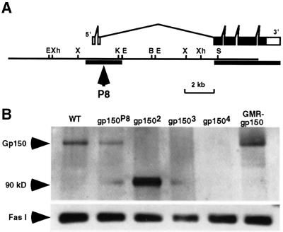
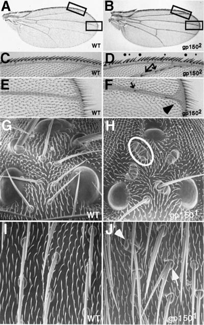
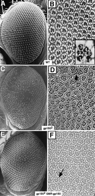
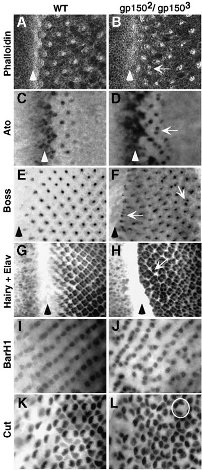
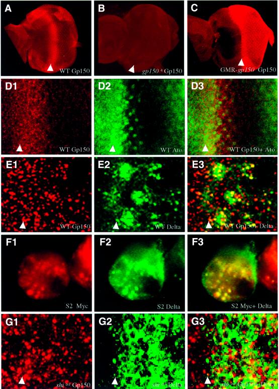
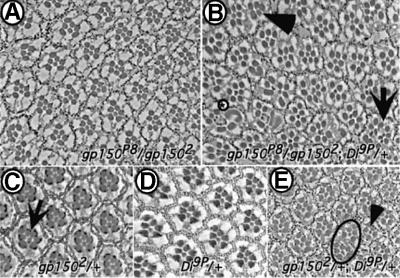
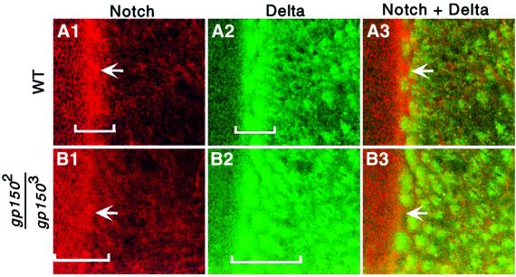
Cellular signaling activities must be tightly regulated for proper cell fate control and tissue morphogenesis. Here we report that the Drosophila leucine-rich repeat transmembrane glycoprotein Gp150 is required for viability, fertility and development of the eye, wing and sensory organs. In the eye, Gp150 plays a critical role in regulating early ommatidial formation. Gp150 is highly expressed in cells of the morphogenetic furrow (MF) region, where it accumulates exclusively in intracellular vesicles in an endocytosis-independent manner. Loss of gp150 function causes defects in the refinement of photoreceptor R8 cells and recruitment of other cells, which leads to the formation of aberrant ommatidia. Genetic analyses suggest that Gp150 functions to modulate Notch signaling. Consistent with this notion, Gp150 is co-localized with Delta in intracellular vesicles in cells within the MF region and loss of gp150 function causes accumulation of intracellular Delta protein. Therefore, Gp150 might function in intracellular vesicles to modulate Delta-Notch signaling for cell fate control and tissue morphogenesis.


Figures

None
Fig. 1. Identification of loss-of-function alleles of the gp150 gene. (A) The gp150 gene consists of six exons and five introns with the complete coding sequence restricted within a 4 kb genomic region. The filled boxes represent a complete 3156 bp ORF. Sequences of two genomic regions indicated by the thick horizontal lines were determined to illustrate intron–exon structures of the gp150 transcriptional unit. A PCR method was used to obtain the downstream genomic sequence of gp150 that was not included in the genomic clone. In P8, a P transposon was inserted in the second intron of gp150. Restriction enzymes used for mapping include BamHI (B), EcoRI (E), KpnI (K), SalI (S), XbaI (X) and XhoI (Xh). (B) A western blot was probed with a Gp150 antibody that was made against the N-terminal region (amino acids 5–192) of Gp150. Since no Gp150 protein was detectable in gp1502, gp1503 and gp1504 mutants, these alleles can be considered as molecular nulls. The gp1502 mutation results in the production of a non-functional 90 kDa protein. This is based on observations that gp1502 is recessive and the mutant phenotypes caused by gp1502 are similar to that of gp1503 and gp1504. The 70 kDa Fas I protein served as an internal control and was detected by anti-Fas I (MAb6D8) antibody in all lanes.
None
Fig. 2. Loss of gp150 function results in defective wing and sensory organs. Wild-type (A, C and E) and gp1502 mutant (B, D and F) wings are shown. (C) and (E) are enlarged from boxed areas in (A). (D) and (F) are enlarged from the boxed areas in (B). Some bristles are missing (dots) or duplicated (asterisks) in the wing margin (D). Arrows point to some ectopic bristles in the second and third veins (D and F) and the arrowhead identifies an enlarged ‘Delta-like’ vein structure (F). SEM images of wild-type (G and I) and gp1501 mutant flies (H and J) are shown to illustrate phenotypes in ocellar bristles (G and H) and sensory organs over the notum (I and J). Multiple socket cells are often developed in sensory organs (circled in H and indicated by an arrowhead in J). The external shaft is often missing or duplicated (indicated by an arrow in J). Moreover, there are cases where both socket and shaft cells are absent in some sensory organs (H). All other gp150 alleles exhibit similar phenotypes in the wing and sensory organs.
None
Fig. 3. Loss of gp150 function causes defective eye development. SEM images (A, C and E) and tangential sections (B, D and F) of adult eyes are presented. (A and B) Wild type. The inset highlights seven R cells arranged in a trapezoidal configuration in each ommatidium and the rhabdomere of R8 cannot be seen in this apical section. (C and D) gp1502 mutants. About 35% (n = 522) of the ommatidia contain either too many or too few R cells. The arrow indicates a fused ommatidium. At basal levels, ommatidia with multiple R8-like cells were observed (data not shown). Other gp150 alleles exhibit similar phenotypes. (E and F) In gp1502 GMR-gp150 flies, overexpression of gp150 in the eye effectively rescues the mutant eye phenotype, with >93% (n = 368) of the ommatidia containing a normal complement of R cells. The arrow indicates a mutant ommatidium occasionally seen in this genotype. Anterior is to the left, except (D), in which anterior might be to the top.
None
Fig. 4. Loss of gp150 function causes defects in early ommatidial development. Wild-type (A, C, E, G, I and K) and gp1502/gp1503 mutant (B, D, F, H, J and L) third instar eye discs are shown. (A and B) Phalloidin staining. (C and D) Anti-Ato antibody staining. Arrows in (B and D) point to closely located ommatidia. (E and F) Anti-Boss antibody staining. Arrows indicate ommatidia containing more than one Boss-positive R8 cells. (G and H) Double staining for Hairy and Elav. The arrow points to an oversized ommatidium near the furrow. (I and J) Anti-BarH1 antibody staining. (K and L) Anti-Cut antibody staining. The circle indicates an ommatidium missing a cone cell. Arrowheads identify the location of the MF in (A–H). Anterior is to the left in all panels.
None
Fig. 5. gp150 is expressed at high levels in the MF region in the developing eye. (A) High levels of Gp150 protein were detected in the MF region of wild-type third instar eye discs. Posterior to the furrow, relatively lower levels of Gp150 can be detected around each individual ommatidium. (B) Gp150 expression was not detectable in gp1504 eye discs. (C) High levels of Gp150 expression were detected in all cells behind the furrow in GMR-gp150 eye discs. (D13) Double-staining reveals Gp150 (red, D1) and Ato (green, D2) expression in eye discs. A superimposed image from (D1) and (D2) is shown in (D3). (E13) Gp150 and Dl proteins were shown to co-localize in subcellular vesicles in the furrow region. (F13) In S2 cells co-transfected with pmt-gp150-myc and pmt-Dl, Gp150 and Dl were also shown to co-localize in subcellular vesicles. (G13) Heat-treated shits1 mutant third instar eye discs were double stained with anti-Gp150 (red in G1) and anti-Dl (green in G2) antibodies. A superimposed image is shown in (G3). Arrowheads indicate the location of the MF, and anterior is to the left in all panels.
None
Fig. 6. Genetic interactions between gp150 and Delta. Tangential sections of adult eyes are presented. (A) The gp150P8/gp1502 eye exhibits ∼19% (n = 578) mutant ommatidia. (B) Reduction of Dl function strongly enhances the gp150 mutant phenotypes. Up to 68% (n = 544) abnormal ommatidia, which contain more R cells (arrow), fewer R cells (arrowhead) or apparently fused ommatidial units, were found in gp150P8/gp1502; Dl9P/+ flies. Often rhabdomeres exhibit abnormal morphology (circled asterisk). (Cgp1502/+. The arrow points to an occasionally observed aberrant ommatidium. (DDl9P/+ flies, no mutant ommatidia can be found. (Egp1502/+; Dl9P/+. The circle indicates a fused ommatidium and the arrowhead points to an ommatidium missing an outer R cell.
None
Fig. 7. Expression of Delta and Notch in wild-type and gp150 mutant eye discs. Wild-type (A13) and gp1502/gp1503 mutant (B13) third instar eye discs were stained with anti-N (red in A1 and B1) and anti-Dl (green in A2 and B2) antibodies. Superimposed images are shown in (A3 and B3). Brackets indicate areas that show relatively higher N or Dl levels in the MF region. Arrows point to N-positive cells in between Dl-positive clusters in the MF. Anterior is to the left in all panels.

Similar articles

Cited by

References

    1. Artavanis-Tsakonas S., Rand,M.D. and Lake,R.J. (1999) Notch signaling: cell fate control and signal integration in development. Science, 284, 770–776. - PubMed
    1. Baker N.E. and Zitron,A.E. (1995) Drosophila eye development: Notch and Delta amplify a neurogenic pattern conferred on the morphogenic furrow by scabrous. Mech. Dev., 49, 173–189. - PubMed
    1. Baker N.E. and Yu,S.-Y. (1997) Proneural function of neurogenic genes in the developing Drosophila eye. Curr. Biol., 7, 122–132. - PubMed
    1. Baker N.E. and Yu,S.-Y. (1998) The R8-photoreceptor equivalence group in Drosophila: fate choice precedes regulated Delta transcription and is independent of Notch gene dose. Mech. Dev., 74, 3–14. - PubMed
    1. Baker N.E., Mlodzik,M. and Rubin,G.M. (1990) Spacing differentiation in the developing Drosophila eye: a fibrinogen-related lateral inhibitor encodes by scabrous. Science, 250, 1370–1377. - PubMed

Publication types

MeSH terms