Comparative Study
. 2002 Mar 1;21(5):1177-87.
doi: 10.1093/emboj/21.5.1177.

The mRNA export factor Dbp5 is associated with Balbiani ring mRNP from gene to cytoplasm

Affiliations
Comparative Study

The mRNA export factor Dbp5 is associated with Balbiani ring mRNP from gene to cytoplasm

Jian Zhao et al. EMBO J. .

Abstract

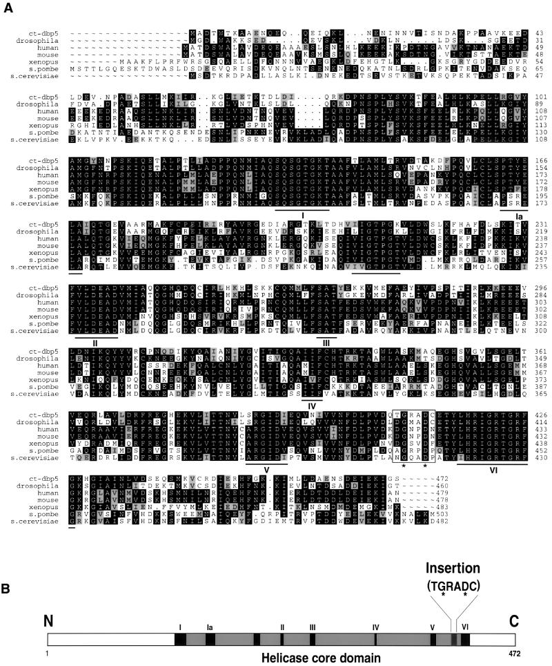
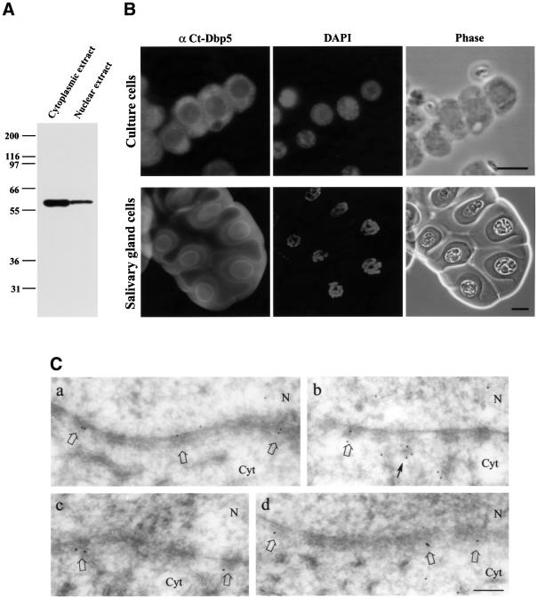
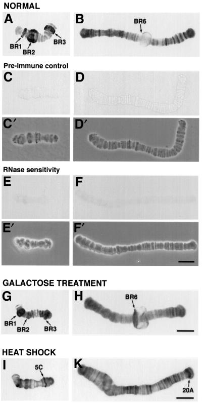
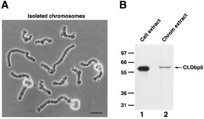
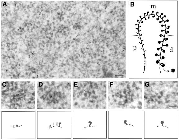
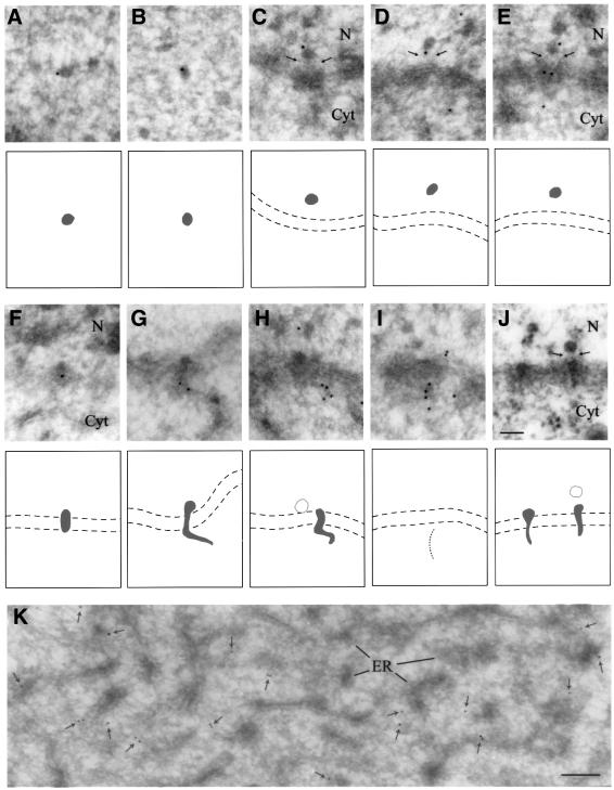
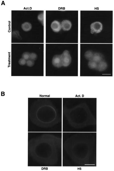
The DEAD box RNA helicase Dbp5 is essential for nucleocytoplasmic transport of mRNA-protein (mRNP) complexes. Dbp5 is present mainly in the cytoplasm and is enriched at the cytoplasmic side of nuclear pore complexes (NPCs), suggesting that it acts in the late part of mRNP export. Here, we visualize the assembly and transport of a specific mRNP particle, the Balbiani ring mRNP in the dipteran Chironomus tentans, and show that a Dbp5 homologue in C.tentans, Ct-Dbp5, binds to pre-mRNP co-transcriptionally and accompanies the mRNP to and through the nuclear pores and into the cytoplasm. We also demonstrate that Ct-Dbp5 accumulates in the nucleus and partly disappears from the NPC when nuclear export of mRNA is inhibited. The fact that Ct-Dbp5 is present along the exiting mRNP fibril extending from the nuclear pore into the cytoplasm supports the view that Ct-Dbp5 is involved in restructuring the mRNP prior to translation. Finally, the addition of the export factor Dbp5 to the growing transcript highlights the importance of the co-transcriptional loading process in determining the fate of mRNA.


Figures

None
Fig. 1. Sequence characteristics of Ct-Dbp5. (A) The amino acid sequence of Ct-Dbp5 compared with Dbp5 in different organisms. Amino acid residues that are identical in at least three species at a given position are shown in white against a black background. Residues that are functionally similar to the predominant one at a position are highlighted against a grey background. The conserved DEAD box helicase motifs are underlined, and the Dbp5-specific insert is boxed. (B) Schematic presentation of Ct-Dbp5. The helicase core domain with its conserved motifs and the Dbp5-specific insert are pointed out. In the insert, the conserved second and fifth positions are marked with a star, in both (A) and (B).
None
Fig. 2. Immunological detection of Ct-Dbp5. (A) Western blot analysis of Ct-Dbp5 in cytoplasmic and nuclear extracts from C.tentans tissue culture cells. (B) Localization of Ct-Dbp5 by immunofluorescence microscopy in C.tentans tissue culture cells (top) and salivary gland cells (bottom). The same preparations are also shown stained with DAPI and in phase contrast. (C) Immunoelectron microscopic localization of Ct-Dbp5 in the vicinity of the nuclear envelope. Labelled NPCs are indicated by open arrows. A cytoplasmic cluster of gold particles close to an NPC is indicated by a thin arrow. N, nucleus; Cyt, cytoplasm. Bar equals 10 µm in (B, upper), 20 µm in (B, lower) and 200 nm in (C).
None
Fig. 3. Immunological detection of Ct-Dbp5 in polytene chromosomes. Polytene chromosomes were isolated from untreated (AF), galactose-treated (G and H) and heat-shocked (I and K) salivary glands and subjected to immunocytology. Some of the untreated chromosomes served as pre-immune controls (C and D), while others were RNase treated (E and F). (C′), (D′), (E′) and (F′), phase contrast images; (A), (C), (E), (G) and (I), chromosome IV; (B) and (H), chromosome III; (D), (F) and (K), chromosome I. After heat shock, the BR genes are switched off and heat shock genes, e.g. in region 5C on chromosome IV (I) and region 20A on chromosome I (K), are turned on. Bar, 20 µm.
None
Fig. 4. The presence of Ct-Dbp5 on isolated polytene chromosomes. (A) Isolated polytene chromosomes from C.tentans salivary gland cells. There are four different chromosomes, two of which carry a nucleolus. Bar, 40 µm. (B) Western blot analysis of extract from the isolated chromosomes. An extract from C.tentans tissue culture cells was used as control.
None
Fig. 5. Immunoelectron microscopic localization of Ct-Dbp5 along transcriptionally active BR genes. (A) Section through a Balbiani ring showing a large number of segments of active genes. Arrows indicate the position of gold particles. (B) Schematic drawing of an active BR gene; growing RNP particles in the promoter-proximal (p), middle (m) and promoter-distal (d) portions of the gene are shown as well as the chromatin axis. (CG) Immunolabelled gene segments at higher magnification; (C) proximal portion; (D) middle portion; (E and F) distal portions. Schematic drawings are shown below the electron micrographs. Labelled, growing RNP particles are depicted in black and the putative chromatin axis by a broken line. Gold particles at the very tip of growing RNP particles are indicated by arrows in (E) and (F). Bar equals 200 nm in (A) and 100 nm in (G).
None
Fig. 6. Immunoelectron microscopic localization of Ct-Dbp5 in nucleoplasmic and translocating BR mRNP particles and in cytoplasm. (A and B) Nucleoplasmic BR RNP particles. (CE) BR particles bound to the top of the nuclear basket; the basket is indicated by thin arrows. (FH) BR particles in various stages of translocation through the central channel of the NPC. In (G) and (H) the exiting RNP fibril is extending into the cytoplasm. (I) A line of gold particles extending from an NPC, presumably indicating the presence of an exiting mRNP fibril. (J) Plastic-embedded, translocating BR particles. The arrows indicate the position of the basket. The two membranes of the nuclear envelope are indicated by broken lines in (C–J). (K) Cytoplasm. The gold particles are indicated by small arrows. N, nucleus; Cyt, cytoplasm; ER, endoplasmic reticulum. Bar equals 100 nm in (J) and 200 nm in (K).
None
Fig. 7. Redistribution of Ct-Dbp5 after treatment with actinomycin D, DRB or heat shock. (AC.tentans tissue culture cells were exposed to actinomycin D, DRB or heat shock and stained with the anti-Ct-Dbp5 antibodies before and after the treatments. Bar equals 10 µm. (BC.tentans salivary gland cells were exposed to actinomycin D, DRB or heat shock, treated briefly with detergents to allow antibodies to enter the cytoplasm, but not the nucleus, and stained with the anti-Ct-Dbp5 antibodies. Bar, 20 µm.

Similar articles

Cited by

References

    1. Alzhanova-Ericsson A.T., Sun X., Visa,N., Kiseleva,E., Wurtz,T. and Daneholt,B. (1996) A protein of the SR family of splicing factors binds extensively to exonic Balbiani ring pre-mRNA and accompanies the RNA from the gene to the nuclear pore. Genes Dev., 10, 2881–2893. - PubMed
    1. Bachi A. et al. (2000) The C-terminal domain of TAP interacts with the nuclear pore complex and promotes export of specific CTE-bearing RNA substrates. RNA, 6, 136–158. - PMC - PubMed
    1. Baurén G. and Wieslander,L. (1994) Splicing of Balbiani ring 1 gene pre-mRNA occurs simultaneously with transcription. Cell, 76, 183–192. - PubMed
    1. Baurén G., Jiang,W.-Q., Bernholm,K., Gu,F. and Wieslander,L. (1996) Demonstration of a dynamic, transcription-dependent organization of pre-mRNA splicing factors in polytene nuclei. J. Cell Biol., 133, 929–941. - PMC - PubMed
    1. Cole C.N. (2000) mRNA export: the long and winding road. Nature Cell Biol., 2, E55–E58. - PubMed

Publication types

MeSH terms