. 2002 Mar 5;99(5):3252-7.
doi: 10.1073/pnas.042701199. Epub 2002 Feb 26.

Visualization of functionally activated circuitry in the brain

Affiliations

Visualization of functionally activated circuitry in the brain

Yvette Wilson et al. Proc Natl Acad Sci U S A. .

Abstract

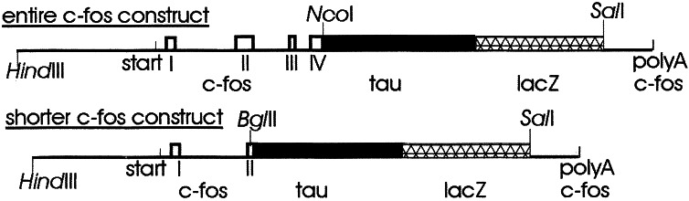
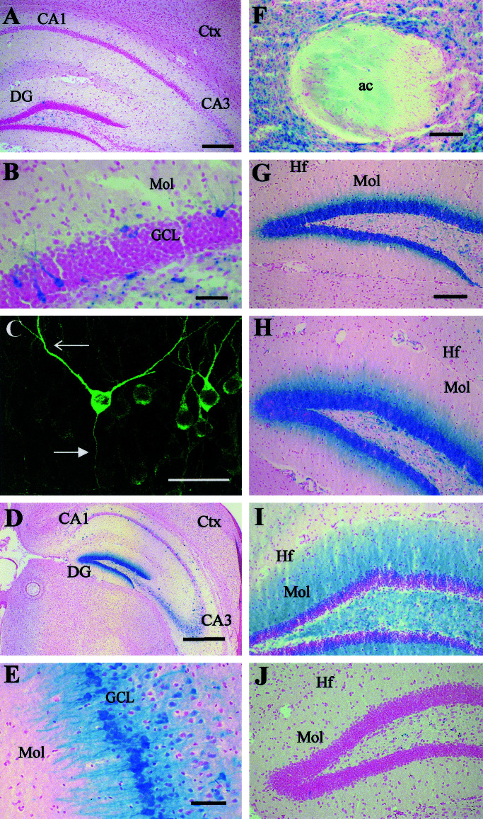
We have used a transgenic approach to visualize functionally activated neurons and their projections. The transgenic mice contain a tau-lacZ fusion gene regulated by the promoter for c-fos, an immediate early gene that is rapidly induced in neurons after functional stimulation. Constitutive expression of beta-galactosidase (beta-gal), the lacZ product, was low and in accord with previous reports of c-fos expression. However, expression of beta-gal in positive neurons was clearly in cell bodies, axons, and dendrites. Treatment of the mice with kainic acid, a strong inducer of c-fos expression, resulted in high induction of beta-gal. beta-gal was induced in the same defined populations of neurons in the brain as those that express c-fos after kainic acid induction. Furthermore, the pattern of beta-gal expression within the neurons changed over time after kainic acid treatment. Early after kainate treatment, beta-gal was found mainly in cell bodies; at later times, expression extended further along the neuronal processes. This expression pattern is consistent with induction and anterograde transport of the Fos-Tau-beta-gal protein in the neurons. To test whether a functionally activated pathway could be visualized, transgenic mice were deprived of water, which activates nuclei involved in body fluid homeostasis. beta-gal induction was traced in neurons and their processes in the lamina terminalis, in magnocellular neurons of the supraoptic and paraventricular nuclei, and in their projections to the posterior pituitary gland. This strategy allowed the mapping of an activated osmoregulatory pathway. This transgenic approach may have general application in the mapping of functionally activated circuitry in the brain.


Figures

Figure 1
Figure 1
Constructs used for the generation of transgenic mice. Clear boxes represent exons of c-fos retained in the constructs, black indicates tau sequence, and crosshatched box indicates lacZ sequence. 5′ and 3′ sequences are derived from genomic c-fos (pc-fos). The transcription start site is indicated (start), and sequence 5′ to this contains identified transcriptional regulatory sequences of c-fos (8, 9).
Figure 2
Figure 2
Expression of β-gal in FTL mice under basal conditions and after treatment with kainic acid. FTL mice were taken directly from their home cages (A–C) and killed, and coronal sections of brains were processed for β-gal. (A) Low-power view of brain, including hippocampus and cortex. (B) Dentate gyrus, showing β-gal-stained cell bodies with processes extending into the molecular layer. (C) High-power confocal image of dentate gyrus showing immunostained granule cells with labeled dendrites (open arrow) and axons (closed arrow). (D–F) Mice were treated with kainate and processed for β-gal expression after 4 h. (D) Hippocampus, most cells are positive in dentate gyrus. (E) High-power view of CA3 region. (F) Anterior commissure. (G–J) Dentate gyrus of animals treated with kainate for 2 h (G), 4 h (H), 24 h (I), and 48 h (J); note staining in molecular layer. ac, Anterior commissure; Ctx, cortex; DG, dentate gyrus; GCL, granule cell layer; Hf, hippocampal fissure; Mol, molecular layer. [Scale bar: 500 μm (A), 50 μm (B and C), 250 μm (D), 60 μm (E), and 125 μm (F–J).]
Figure 3
Figure 3
Expression of β-gal in FTL mice after water deprivation. (A) Schematic diagram of the osmoregulatory pathway investigated. Neurons (filled red circles) in the lamina terminalis (consisting of the organum vasculosum of the lamina terminalis, median preoptic nucleus, and subfornical organ) that are responsive to plasma hypertonicity send efferent axonal projections (red lines) to magnocellular neurons (filled blue circles) in the supraoptic nucleus and hypothalamic paraventricular nucleus. The processes (blue lines) of these magnocellular neurons form the hypothalamo-neurohypophysial pathway that courses in the median eminence to reach the posterior pituitary, where neurosecretion of vasopressin and oxytocin into the general circulation occurs as the result of this pathway being activated in response to dehydration. Mice were either water replete (C, E, G, and I) or deprived (B, D, F, H, and J) for 48 h and analyzed for β-gal expression by histochemistry (B–H) or immunofluorescence (I and J). (B) High-power view of region of the organum vasculosum of the lamina terminalis indicated with open arrow in D. (C and D) Sagittal sections of midline of ventral brain around third ventricle. (E and F) Hypothalamus, showing the SON and PVN. (G and H) Sections through the median eminence. (I and J) Sections through the pituitary. Ten mice from control and water-deprived animals were analyzed, and figures are representative of findings from each of the animals. 3V, third ventricle; ∗, anterior commissure; AL, IL, and PL, anterior, intermediate, and posterior lobe of pituitary, respectively; iME, internal median eminence; MnPO, median preoptic nucleus; oc, optic chiasm; ot, optic tract; OVLT, organum vasculosum of the lamina terminalis; Pit, pituitary; PVN, hypothalamic paraventricular nucleus. [Scale bar: 50 μm (B), 500 μm (C and D), 200 μm (E and F), and 100 μm (G–J).]

References

    1. Morgan J I, Curran T. Annu Rev Neurosci. 1991;14:421–451. - PubMed
    1. Herdegen T, Leah J D. Brain Res Rev. 1998;28:370–490. - PubMed
    1. Herrera D G, Robertson H A. Prog Neurobiol. 1996;50:83–107. - PubMed
    1. Chaudhuri A. NeuroReport. 1997;8:v–ix. - PubMed
    1. Callahan C A, Thomas J B. Proc Natl Acad Sci USA. 1994;91:5972–5976. - PMC - PubMed

Publication types

MeSH terms

LinkOut - more resources