

doi: 10.1073/pnas.052005999.
Epub 2002 Feb 26.
Network of coupled promoting motions in enzyme catalysis
Affiliations
- PMID: 11867722
- PMCID: PMC122427
- DOI: 10.1073/pnas.052005999
Item in Clipboard
Network of coupled promoting motions in enzyme catalysis
Proc Natl Acad Sci U S A.
.
Abstract
A network of coupled promoting motions in the enzyme dihydrofolate reductase is identified and characterized. The present identification is based on genomic analysis for sequence conservation, kinetic measurements of multiple mutations, and mixed quantum/classical molecular dynamics simulations of hydride transfer. The motions in this network span time scales of femtoseconds to milliseconds and are found on the exterior of the enzyme as well as in the active site. This type of network has broad implications for an expanded role of the protein fold in catalysis as well as ancillaries such as the engineering of altered protein function and the action of drugs distal to the active site.
Figures
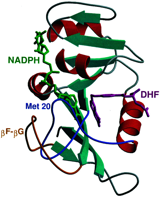
Secondary structure of DHFR. The Met-20 and βF–βG loops, as well
as the NADPH coenzyme and DHF substrate, are labeled. All structural
figures in this paper were generated by using the programs
MOLSCRIPT and RASTER3D.
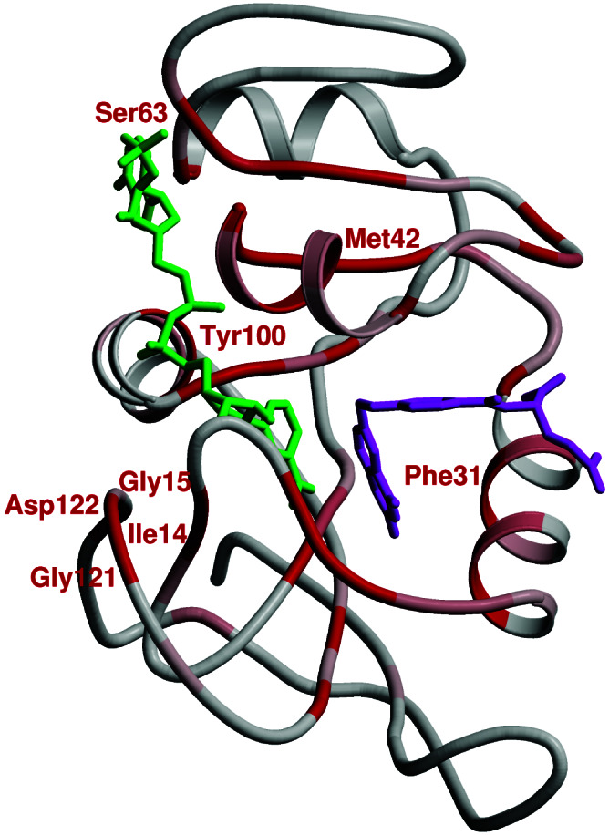
Sequence conservation in DHFR. Regions of conservation are mapped onto
the structure of E. coli DHFR by using a gradient color
scheme (gray to red, where red is the most conserved). NADPH and DHF
are in green and magenta, respectively.
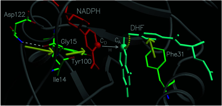
Diagram of a portion of the network of coupled promoting motions in
DHFR. The yellow arrows and arc indicate the coupled promoting
motions.
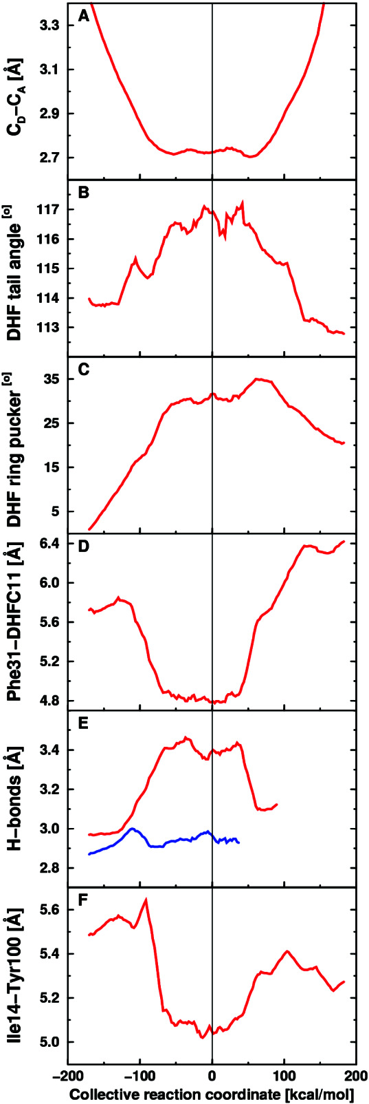
Equilibrium averages of geometrical properties along the collective
reaction coordinate. (A) Donor–acceptor distance.
(B) Angle between the acceptor and methylene amino
linkage in DHF. (C) DHF pterin ring puckering angle.
(D) Distance between Cζ of Phe-31 and C11
of DHF. (E) Hydrogen-bonding distance between N of
Asp-122 and O of Gly-15 (red) and between O of Ile-14 and
carboxamide N of NADPH (blue). (F) Distance between
Cδ of Ile-14 and the side-chain oxygen of Tyr-100.
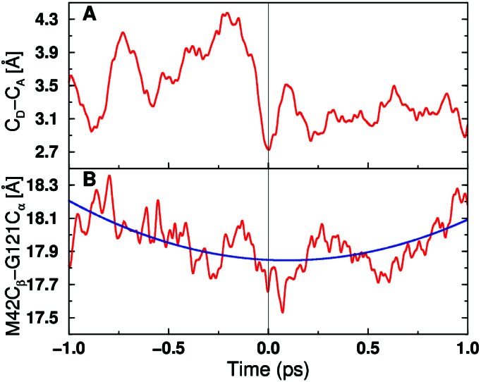
Time evolution of two select distances for a representative real-time
vibrationally adiabatic trajectory. (A) Donor-acceptor
distance. (B) Distance between Cα of
Gly-121 and Cβ of Met-42. Time t = 0
corresponds to the transition state, and the reaction evolves toward
the reactant/product as the time becomes negative/positive. For
B, the blue curve indicates a fit to a frequency on the
picosecond time scale.
References
-
- Hammes G G. Nature (London) 1964;204:342–343. - PubMed
-
- Kohen A, Cannio R, Bartolucci S, Klinman J P. Nature (London) 1999;399:496–499. - PubMed
-
- Karplus M. J Phys Chem B. 2000;104:11–27.
-
- Osborne M J, Schnell J, Benkovic S J, Dyson H J, Wright P E. Biochemistry. 2001;40:9846–9859. - PubMed
-
- Radkiewicz J L, Brooks C L., III J Am Chem Soc. 2000;122:225–231.
Publication types
MeSH terms
Substances
Grants and funding
LinkOut - more resources
Full Text Sources
Other Literature Sources
