. 2002 Mar 5;99(5):2714-9.
doi: 10.1073/pnas.052284899. Epub 2002 Feb 26.

Interaction with glycosaminoglycans is required for cyclophilin B to trigger integrin-mediated adhesion of peripheral blood T lymphocytes to extracellular matrix

Affiliations

Interaction with glycosaminoglycans is required for cyclophilin B to trigger integrin-mediated adhesion of peripheral blood T lymphocytes to extracellular matrix

Fabrice Allain et al. Proc Natl Acad Sci U S A. .

Abstract

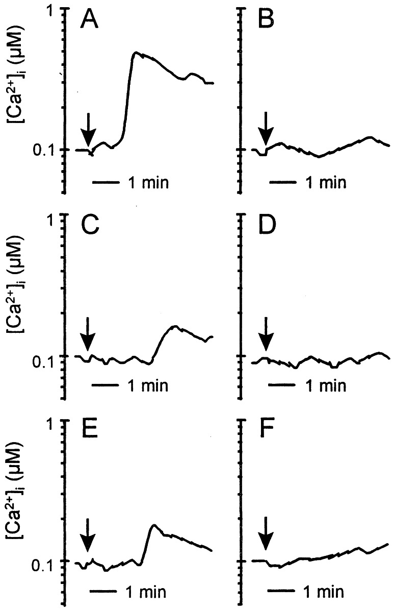
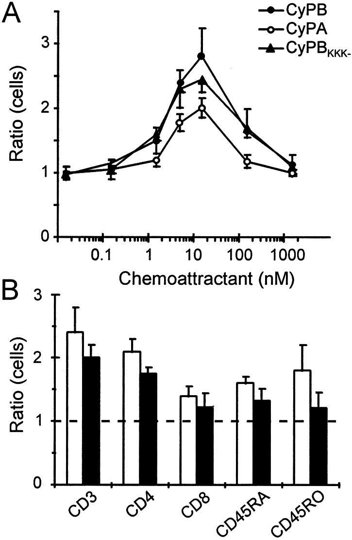
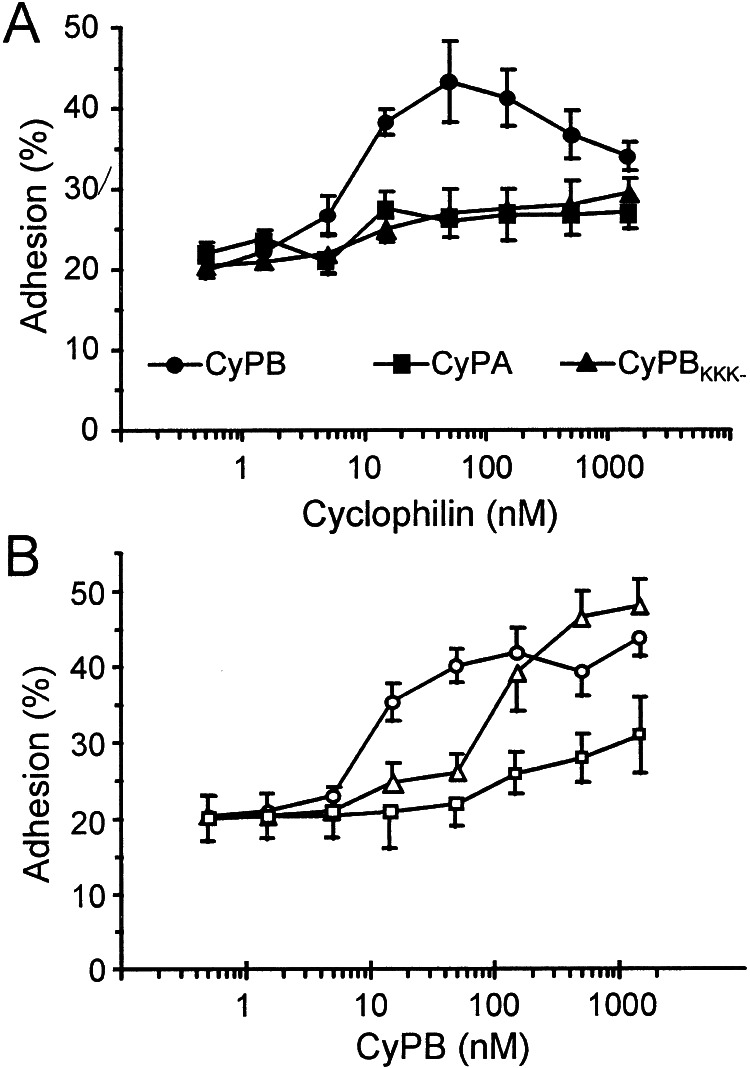
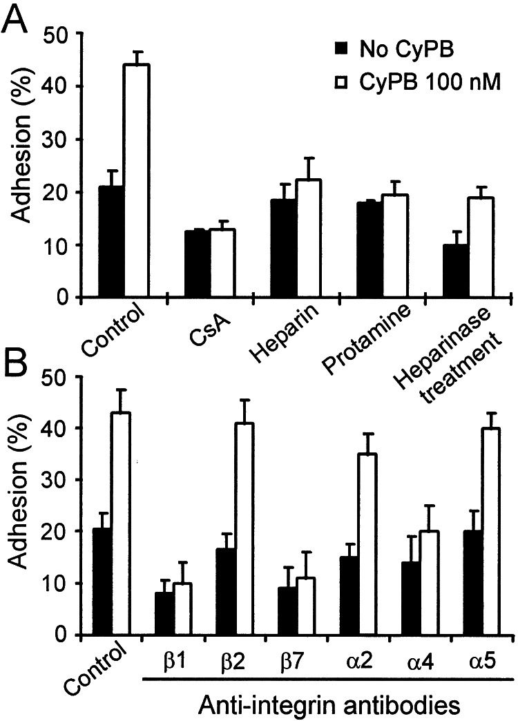
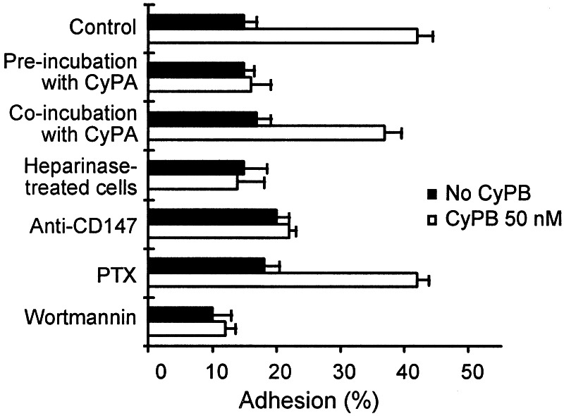
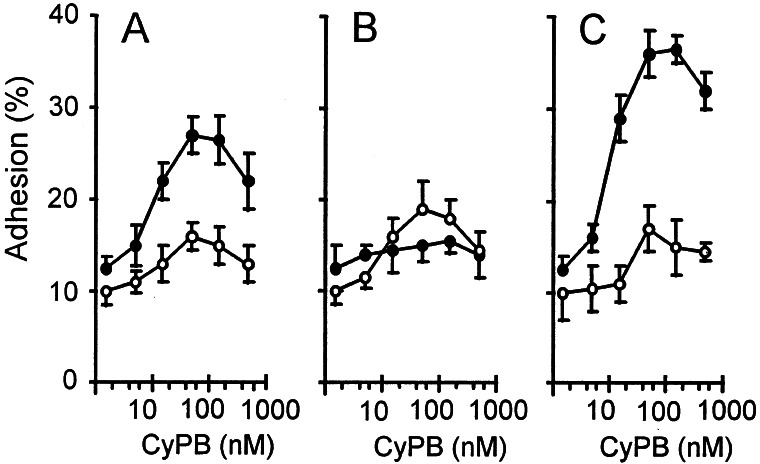
Cyclophilins A and B (CyPA and CyPB) are cyclosporin A-binding proteins that are involved in inflammatory events. We have reported that CyPB interacts with two types of cell-surface-binding sites. The first site corresponds to a functional receptor and requires interaction with the central core of CyPB. This region is highly conserved in cyclophilins, suggesting that CyPA and CyPB might share biological activities mediated by interaction with this receptor. The second site is identified with glycosaminoglycans (GAGs), the binding region located in the N terminus of CyPB. The difference in the N-terminal extensions of CyPA and CyPB suggests that a unique interaction with GAGs might account for selective activity of CyPB. To explore this hypothesis, we analyzed the lymphocyte responses triggered by CyPA, CyPB, and CyPB(KKK-), a mutant unable to interact with GAGs. The three ligands seemed capable enough to elicit calcium signal and chemotaxis by binding to the same signaling receptor. In contrast, only CyPB enhanced firm adhesion of T cells to the extracellular matrix. This activity depended on the interactions with GAGs and signaling receptor. CyPB-mediated adhesion required CD147 presumably because it was a costimulatory molecule and was related to an activation of alpha4beta1 and alpha4beta7 integrins. Finally, we showed that CyPB was capable mainly to enhance T cell adhesion of the CD4+CD45RO+ subset. The present data indicate that CyPB rather than CyPA is a proinflammatory factor for T lymphocytes and highlight the crucial role of CyPB-GAG interaction in the chemokine-like activity of this protein.


Figures

Figure 1
Figure 1
Cyclophilin-mediated Ca2+ signaling in T lymphocytes. Ca2+ mobilization in Fluo-3-loaded T cells was measured in the presence of CyPB (50 nM) (A and B), CyPA (250 nM) (C and D), CyPBKKK− (50 nM) (E), or CyPB/CsA (50 nM) (F). The arrows indicate the addition of the agonist. Cells were either directly stimulated with the agonist for analysis (A, C, E, F) or prestimulated with CyPA (B) or CyPB (D) before a second stimulation. Changes in fluorescence, reflecting changes in cytosolic Ca2+ concentration, were monitored by flow cytofluorimetry. Tracings are from a single representative experiment.
Figure 2
Figure 2
Chemotactic activity of CyPA and CyPB. Results are expressed as the number of cells migrating toward the test sample divided by the number of cells migrating toward control medium. (A) Comparison of chemotactic activities of CyPB (●), CyPA (○), and CyPBKKK− (▴). Data represent means ± SD of triplicate from a representative experiment. (B) Chemotactic preferential responses of T cell subsets to CyPA (filled bars) or CyPB (open bars). Data are means ± SD from three separate experiments.
Figure 3
Figure 3
Enhancing effect of CyPB on T lymphocyte adhesion to the ECM. (A) Comparison of the effects of CyPB (●), CyPBKKK− (▴), and CyPA (■) on T cell adhesion. T lymphocytes were stimulated in the presence of increasing concentrations of recombinant cyclophilins and allowed to adhere into ECM-coated wells. (B) Effects of anchored or diffusible CyPB on T cell adhesion. In one procedure (Δ), ECM-coated plates were pretreated with CyPB and extensively washed before the addition of T lymphocytes. In the other procedures, T cells were pretreated with CyPB, either at 37°C (○) or at 4°C (□), and then allowed to adhere into ECM-coated wells. Results are expressed as percentages of initially added T lymphocytes (1 × 10⁶ per well) remaining associated to the ECM-coated well. Points represent means ± SD of quadruplicates and are representative from at least three separate experiments.
Figure 4
Figure 4
Inhibition of CyPB-mediated adhesion of peripheral blood T lymphocytes to the ECM. T lymphocytes were preincubated in the presence of inhibitors for 30 min at 37°C and thereafter used for adhesion assays in the absence (filled bars) or the presence of 100 nM CyPB (open bars). (A) Inhibitory properties of CsA (1 μM), heparin (500 μg/ml), protamine (12.5 μM), and treatment of the ECM by heparinase on CyPB-mediated adhesion. (B) Effect of blocking antibodies to integrin subunits on T lymphocyte adhesion to the ECM. Data are means ± SD of quadruplicates from at least three separate experiments.
Figure 5
Figure 5
Involvement of cell-surface proteoglycans and cosignaling molecules in CyPB-mediated adhesion of T lymphocytes to fibronectin. Peripheral blood T cells from the same donors were either treated with heparinase or incubated with CyPA (500 nM), monoclonal antibody to CD147, PTX (2 μg/ml), or wortmannin (50 nM). T lymphocytes were then allowed to adhere into fibronectin-coated wells in the absence (filled bars) or presence of 50 nM CyPB (open bars). Data are means ± SD of quadruplicates from at least three separate experiments.
Figure 6
Figure 6
Phenotype of responsive T cells to CyPB. T lymphocytes were stimulated with increasing concentrations of CyPB and added to fibronectin-coated wells. Adherent cells were fixed and immunostained with either anti-CD4 (●) or anti-CD8 (○) monoclonal antibodies. Discrimination between CD4+ and CD8+ adherent T cells was performed in the whole T cell population (A) or after separation of T lymphocytes within CD45RA+ native (B) and CD45RO+ memory (C) T cell subsets. Data represent means ± SD of quadruplicates and are representative from at least three separate experiments.

References

    1. Handschumacher R E, Harding M W, Rice J, Drug R J, Speicher D W. Science. 1984;226:544–547. - PubMed
    1. Schreiber S L. Science. 1991;251:283–287. - PubMed
    1. Fischer G, Wittmann-Liebold B, Lang K, Kiefhaber T, Schmid F X. Nature (London) 1989;337:476–478. - PubMed
    1. Takahashi N, Hayano T, Suzuki M. Nature (London) 1989;337:473–475. - PubMed
    1. Liu J, Farmer J D, Lane W S, Friedmann J, Weissman I, Schreiber S L. Cell. 1991;66:807–815. - PubMed

Publication types

LinkOut - more resources