. 2002 Jan 21;86(2):274-81.
doi: 10.1038/sj.bjc.6600038.

Identification of molecular markers for the early detection of human squamous cell carcinoma of the uterine cervix

Affiliations
Free PMC article

Identification of molecular markers for the early detection of human squamous cell carcinoma of the uterine cervix

Q Cheng et al. Br J Cancer. .
Free PMC article

Abstract

To identify novel cellular genes that could potentially act as predictive molecular markers for human cervical cancer, we employed RT--PCR differential display, reverse Northern and Northern blot analysis to compare the gene expression profiles between squamous cell carcinoma biopsies and adjacent histo-pathological normal epithelium tissues. Twenty-eight cDNA clones were isolated that were demonstrated to be consistently over-expressed in squamous cell cervical cancer biopsies of FIGO stages 1B to 3B. Most importantly, it was observed that, in addition to their over-expression in cancer lesions, some of these genes are upregulated in the presumably histo-pathological normal adjacent tissues. Of particular interest is clone G30CC that has been identified to be the gene that encodes S12 ribosomal protein. When employed for RNA--RNA in situ hybridization experiments, expression of G30CC could be detected in the immature basal epithelial cells of histo-pathological normal tissues collected from cervical cancer patients of early FIGO stages. In comparison, the expression of G30CC was not detected in cervical tissues collected from patients admitted for surgery of non-malignant conditions. These results allow the distinct possibility of employing the ribosomal protein S12 gene as an early molecular diagnostic identifier for the screening of human cervical cancer and a potential target employed for cancer gene therapy trials.


Figures

Figure 1
Figure 1
Differential display analysis. Purified total RNA from cervical cancer biopsies was reversed transcribed with the primer P21 (see Materials and Methods). The reverse transcribed cDNA were then amplified by PCR in the presence of 1μCi [α-33P]dATP and either primer P30, P31, or P32. The PCR products were then separated on polyacrylamide gel. (A) RT–PCR differential display with the primer pair P21/P30. (B) RT–PCR differential display with the primer pair P21/P31. (C) RT–PCR differential display with the primer pair P21/P32. Seventeen bands selected following differential display analyses with the corresponding primer pairs, as indicated in the figure, were employed for subsequent cloning.
Figure 2
Figure 2
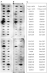
Reverse Northern blot. DNA (2 μg) of the amplified gene fragment of each of the purified clones, along with various amount of α-actin DNA (0.5, 1, 2, or 4 μg) were blotted on Hybond-N+ nylon transfer membrane using the slot-blot apparatus. After denaturation, neutralization and DNA fixation, the filters containing the spotted DNA were hybridized to 32P-labelled cDNA probes. (A) Autoradiogram obtained after Reverse Northern blot hybridization of the spotted cloned gene fragments to a probe prepared by reverse transcription of 30 μg of total RNA purified from the pooled tumour biopsies of six cervical cancer patients. (B) Autoradiogram obtained after Reverse Northern blot hybridization of the spotted cloned gene fragments to a probe prepared by reverse transcription of 30 μg of total RNA purified from the corresponding pooled normal tissue biopsies of the same six cervical cancer employed in (A). (C) The identity and orientation of the cloned gene fragments as spotted on the filter membranes.
Figure 3
Figure 3
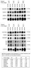
Northern blot analysis employing clones G32C4B, G30CC, G30Ca, G31C5G, G30CI, G32C2B and G31C5E as probes with total RNA obtained from human squamous cell cervical carcinoma biopsies of different FIGO stages and their corresponding adjacent normal tissues. The summary table shows the folds of increase of the cloned cDNA fragments in the various tumour biopsies in comparison to their matched normal counterparts after normalization to G3PDH mRNA.
Figure 4
Figure 4
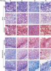
Expression of G30CC in human squamous cell cervical carcinoma of different FIGO stages (1B, 2A, 2B and 3B), as well as their corresponding adjacent normal via RNA–RNA in situ hybridization analysis. The RNA–RNA in situ hybridization was performed by DIG-labelled cRNA probe using the DIG RNA labelling Kit from Boehringer Mannheim. Sub-clones with the G30CC gene fragment inserted in opposite orientation were selected for the synthesis of the sense and anti-sense hybridization probes. Frozen tumour sections (10 μm thick) were sectioned from the frozen tumour biopsies and reacted with the DIG-labelled cRNA probe. After hybridization and washing, the slides were analyzed using the Olympus BX 60 microscope. The histology identifications were performed on consecutive tissue sections following H&E staining. The magnification shown was 600×.
Figure 5
Figure 5
Expression of G30CC in normal epithelium of tissue biopsies obtained from four different non-cervical cancer patients via RNA–RNA in situ hybridization analysis. Sub-clones with the G30CC gene fragment inserted in opposite orientation were selected for the synthesis of the sense and anti-sense hybridization probes. The magnification shown was 600×.

Similar articles

Cited by

References

    1. BassetPBellocqJPWolfCStollIHutinPLimacherJMPodhajcerOLChenardMPRioMCChambonP1990A novel metalloproteinase gene specifically expressed in stromal cells of breast carcinomas Nature 348699704 - PubMed
    1. BosABvan BallegooijenMvan OortmarssenGJvan MarleMEHabbemaJDLyngeE1997Non-progression of cervical intraepithelial neoplasia estimated from population-screening data Br J Cancer 75124130 - PMC - PubMed
    1. BoschFXManosMMMunozNShermanMJansenAMPetoJSchiffmanMHMorenoVKurmanRShahKV1995Prevalence of human papillomavirus in cervical cancer: a worldwide perspective J Natl Cancer Inst 87796802 - PubMed
    1. BurkeTWHoskinsWJHellerPBShenMCWeiserEBParkRC1987Clinical patterns of tumor recurrence after radical hysterectomy in stage IB cervical carcinoma Obstet Gynecol 69382385 - PubMed
    1. ChanseobSZhangWChangHRLeeJH1998Profiling of differentially expressed genes in human primary cervical cancer by complementary DNA expression array Clin Cancer Res 430453050 - PubMed

MeSH terms