

Hypoxia-associated spontaneous pulmonary metastasis in human melanoma xenografts: involvement of microvascular hot spots induced in hypoxic foci by interleukin 8
- PMID: 11870523
- PMCID: PMC2375178
- DOI: 10.1038/sj.bjc.6600052
Hypoxia-associated spontaneous pulmonary metastasis in human melanoma xenografts: involvement of microvascular hot spots induced in hypoxic foci by interleukin 8
Abstract
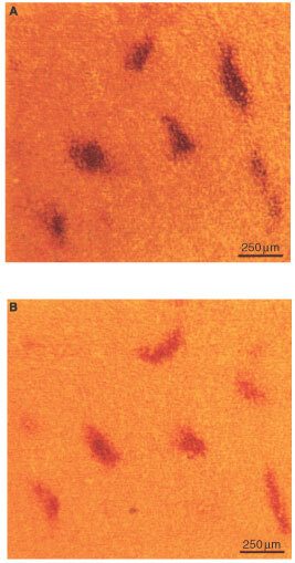
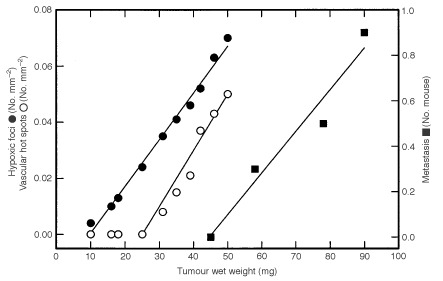
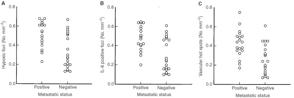
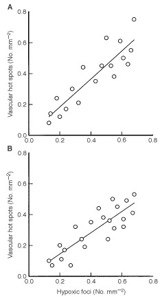
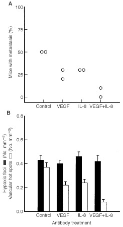
The aim of this study was to investigate whether tumour hypoxia and/or vascular hot spots promote the development of metastatic disease. The D-12 human melanoma xenograft line was used as a tumour model. Hypoxia and vascular hot spots were detected by immunohistochemistry using pimonidazole as a hypoxia marker and anti-CD31 antibody to visualize endothelial cells. Vascular hot spots were found to be induced in hypoxic foci, owing to hypoxia-induced up-regulation of angiogenesis stimulatory factors. This effect was mediated by interleukin 8 and possibly also by vascular endothelial growth factor. Interleukin 8 positive foci showed a high degree of co-localization with hypoxic foci, as revealed by immunohistochemistry. The incidence of spontaneous pulmonary metastases was associated with the density of hypoxic foci, the density of interleukin 8 positive foci and the density of vascular hot spots in the primary tumour. Treatment with neutralizing antibody against interleukin 8 and/or vascular endothelial growth factor resulted in hypoxia-induced necrosis rather than hypoxia-induced vascular hot spots and inhibited metastasis. Our study suggests a cause-effect relationship between hypoxia and metastasis in cancer and hence an elevated probability of metastatic disease in patients having primary tumours characterized by high densities of hypoxic foci and vascular hot spots.
Copyright 2002 The Cancer Research Campaign
Figures

Similar articles
-
Hypoxia-induced metastasis of human melanoma cells: involvement of vascular endothelial growth factor-mediated angiogenesis.Br J Cancer. 1999 Aug;80(11):1697-707. doi: 10.1038/sj.bjc.6690586. Br J Cancer. 1999. PMID: 10468285 Free PMC article.
-
Vascular endothelial growth factor, interleukin 8, platelet-derived endothelial cell growth factor, and basic fibroblast growth factor promote angiogenesis and metastasis in human melanoma xenografts.Cancer Res. 2000 Sep 1;60(17):4932-8. Cancer Res. 2000. PMID: 10987309
-
The tumor bed effect: increased metastatic dissemination from hypoxia-induced up-regulation of metastasis-promoting gene products.Cancer Res. 2005 Mar 15;65(6):2387-96. doi: 10.1158/0008-5472.CAN-04-3039. Cancer Res. 2005. PMID: 15781654
-
Angiogenesis, lymphangiogenesis, and melanoma metastasis.Oncogene. 2003 May 19;22(20):3172-9. doi: 10.1038/sj.onc.1206457. Oncogene. 2003. PMID: 12789293 Review.
-
Angiogenesis and cancer metastasis: antiangiogenic therapy of human pancreatic adenocarcinoma.Int J Clin Oncol. 2001 Apr;6(2):59-65. doi: 10.1007/pl00012085. Int J Clin Oncol. 2001. PMID: 11706751 Review. No abstract available.
Cited by
-
Comparative expression pathway analysis of human and canine mammary tumors.BMC Genomics. 2009 Mar 27;10:135. doi: 10.1186/1471-2164-10-135. BMC Genomics. 2009. PMID: 19327144 Free PMC article.
-
Computer simulation of glioma growth and morphology.Neuroimage. 2007;37 Suppl 1(Suppl 1):S59-70. doi: 10.1016/j.neuroimage.2007.03.008. Epub 2007 Mar 23. Neuroimage. 2007. PMID: 17475515 Free PMC article.
-
Lenalidomide modulates IL-8 and anti-prostate antibody levels in men with biochemically recurrent prostate cancer.Prostate. 2012 Apr;72(5):487-98. doi: 10.1002/pros.21449. Epub 2011 Jul 11. Prostate. 2012. PMID: 21748755 Free PMC article. Clinical Trial.
-
Temporal heterogeneity in oxygen tension in human melanoma xenografts.Br J Cancer. 2003 Jul 21;89(2):350-6. doi: 10.1038/sj.bjc.6601047. Br J Cancer. 2003. PMID: 12865929 Free PMC article.
-
Metastasis in melanoma xenografts is associated with tumor microvascular density rather than extent of hypoxia.Neoplasia. 2010 Nov;12(11):889-98. doi: 10.1593/neo.10712. Neoplasia. 2010. PMID: 21076614 Free PMC article.
References
-
- Bar-EliM1999Role of interleukin-8 in tumor growth and metastasis of human melanoma Pathobiology 671218 - PubMed
-
- BrizelDMScullyPSHarrelsonJMLayfieldLJBeanJMProsnitzLRDewhirstMW1996Tumor oxygenation predicts for the likelihood of distant metastases in human soft tissue sarcoma Cancer Res 56941943 - PubMed
-
- FerraraNDavis-SmythT1997The biology of vascular endothelial growth factor Endocr Rev 18425 - PubMed
-
- FidlerIJEllisLM1994The implications of angiogenesis for the biology and therapy of cancer metastasis Cell 79185188 - PubMed
Publication types
MeSH terms
Substances
LinkOut - more resources
Full Text Sources
Other Literature Sources
Medical
