

Combined MR proton lung perfusion/angiography and helium ventilation: potential for detecting pulmonary emboli and ventilation defects
- PMID: 11870828
- PMCID: PMC2230619
- DOI: 10.1002/mrm.10091
Combined MR proton lung perfusion/angiography and helium ventilation: potential for detecting pulmonary emboli and ventilation defects
Abstract
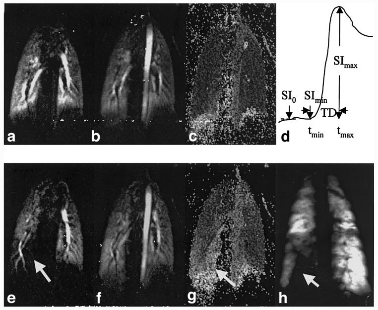
Three-dimensional (3D) perfusion imaging allows the assessment of pulmonary blood flow in parenchyma and main pulmonary arteries simultaneously. MRI using laser-polarized (3)He gas clearly shows the ventilation distribution with high signal-to-noise ratio (SNR). In this report, the feasibility of combined lung MR angiography, perfusion, and ventilation imaging is demonstrated in a porcine model. Ultrafast gradient-echo sequences have been used for 3D perfusion and angiographic imaging, in conjunction with the use of contrast agent injections. 2D multiple-section (3)He imaging was performed subsequently by inhalation of 450 ml of hyperpolarized (3)He gas. The MR techniques were examined in a series of porcine models with externally delivered pulmonary emboli and/or airway occlusions. With emboli, perfusion deficits without ventilation defects were observed; airway occlusion resulted in matched deficits in perfusion and ventilation. High-resolution MR angiography can unambiguously reveal the location and size of the blood emboli. The combination of the three imaging methods may provide complementary information on abnormal lung anatomy and function.
Copyright 2002 Wiley-Liss, Inc.
Figures
Similar articles
-
Feasibility of combining MR perfusion, angiography, and 3He ventilation imaging for evaluation of lung function in a porcine model.Acad Radiol. 2005 Feb;12(2):202-9. doi: 10.1016/j.acra.2004.11.021. Acad Radiol. 2005. PMID: 15721597 Free PMC article.
-
Pulmonary embolism: comprehensive evaluation with MR ventilation and perfusion scanning with hyperpolarized helium-3, arterial spin tagging, and contrast-enhanced MRA.J Vasc Interv Radiol. 2005 Jul;16(7):999-1005. doi: 10.1097/01.RVI.0000162416.64412.FC. J Vasc Interv Radiol. 2005. PMID: 16002508
-
Laser-polarized (3)He as a probe for dynamic regional measurements of lung perfusion and ventilation using magnetic resonance imaging.Magn Reson Med. 2000 Jul;44(1):1-4. doi: 10.1002/1522-2594(200007)44:1<1::aid-mrm1>3.0.co;2-u. Magn Reson Med. 2000. PMID: 10893513
-
Hyperpolarized helium-3 gas magnetic resonance imaging of the lung.Top Magn Reson Imaging. 2003 Jun;14(3):223-30. doi: 10.1097/00002142-200306000-00002. Top Magn Reson Imaging. 2003. PMID: 12973129 Review.
-
Functional MR imaging of pulmonary ventilation using hyperpolarized noble gases.Acta Radiol. 2000 Nov;41(6):519-28. doi: 10.1080/028418500127346009. Acta Radiol. 2000. PMID: 11092469 Review.
Cited by
-
Feasibility of combining MR perfusion, angiography, and 3He ventilation imaging for evaluation of lung function in a porcine model.Acad Radiol. 2005 Feb;12(2):202-9. doi: 10.1016/j.acra.2004.11.021. Acad Radiol. 2005. PMID: 15721597 Free PMC article.
-
In vivo methods and applications of xenon-129 magnetic resonance.Prog Nucl Magn Reson Spectrosc. 2021 Feb;122:42-62. doi: 10.1016/j.pnmrs.2020.11.002. Epub 2020 Dec 9. Prog Nucl Magn Reson Spectrosc. 2021. PMID: 33632417 Free PMC article. Review.
-
Methods of Prospective Investigation of Pulmonary Embolism Diagnosis III (PIOPED III).Semin Nucl Med. 2008 Nov;38(6):462-70. doi: 10.1053/j.semnuclmed.2008.06.003. Semin Nucl Med. 2008. PMID: 19331840 Free PMC article. Review.
-
Pulmonary perfusion imaging in the rodent lung using dynamic contrast-enhanced MRI.Magn Reson Med. 2008 Feb;59(2):289-97. doi: 10.1002/mrm.21353. Magn Reson Med. 2008. PMID: 18228577 Free PMC article.
-
Improving temporal resolution of pulmonary perfusion imaging in rats using the partially separable functions model.Magn Reson Med. 2010 Oct;64(4):1162-70. doi: 10.1002/mrm.22500. Magn Reson Med. 2010. PMID: 20564601 Free PMC article.
References
-
- Hatabu H, Alsop D, Bonnet M, Listerud J, Pietra G, Gefter W. Approaches to MR imaging of lung parenchyma utilizing ultrashort TE gradient echo and fast SE sequences; Proc 2nd Annual Meeting SMR; San Francisco. 1994.p. 1474.
-
- Alsop DC, Hatabu H, Bonnet M, Listerud J, Gefter W. Multi-slice, breathhold imaging of the lung with submillisecond echo times. Magn Reson Med. 1995;33:678–682. - PubMed
-
- Hatabu H, Gaa J, Kim D, Li W, Prasad PV, Edelman RR. Pulmonary perfusion: qualitative assessment with dynamic contrast-enhanced MRI using ultra-short TE and inversion recovery turbo FLASH. Magn Reson Med. 1996;36:503–508. - PubMed
-
- Berthezene Y, Croisille P, Wiart M, Howarth N, Houzard C, Faure O, Douek P, Amiel M, Revel D. Prospective comparison of MR lung perfusion and lung scintigraphy. J Magn Reson Imag. 1999;9:61–68. - PubMed
-
- Schoenberg SO, Bock M, Floemer F, Grau A, Williams DM, Laub G, Knopp MV. High-resolution pulmonary arterio- and venography using multiple-bolus multiphase 3D-Gd-mRA. J Magn Reson Imag. 1999;10:339–346. - PubMed
Publication types
MeSH terms
Substances
Grants and funding
LinkOut - more resources
Full Text Sources
Other Literature Sources
Medical
