

Mycobacterial antigens induce apoptosis in human purified protein derivative-specific alphabeta T lymphocytes in a concentration-dependent manner
- PMID: 11872098
- PMCID: PMC1782647
- DOI: 10.1046/j.0019-2805.2001.01355.x
Mycobacterial antigens induce apoptosis in human purified protein derivative-specific alphabeta T lymphocytes in a concentration-dependent manner
Abstract
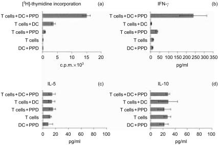
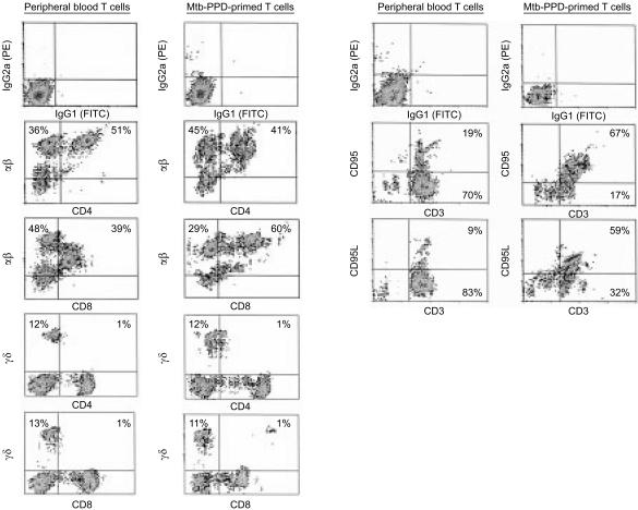
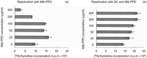
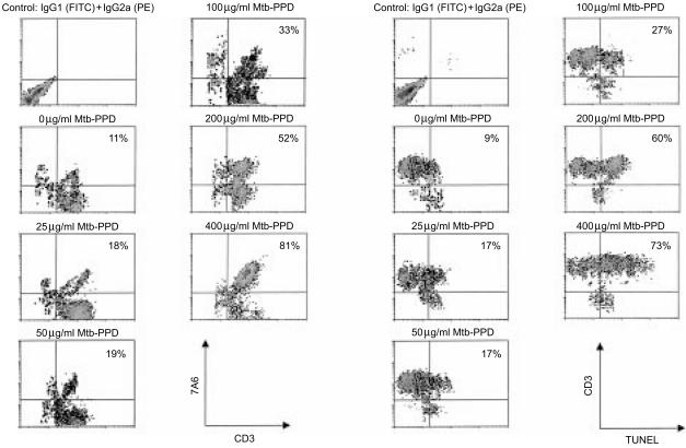
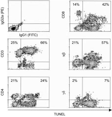
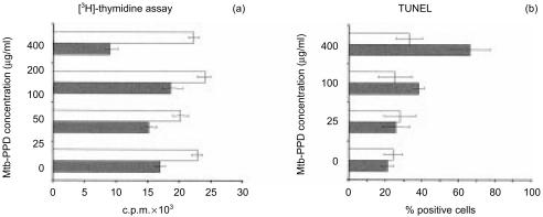
The morbidity and lethality of tuberculosis is partially the result of an ineffective delayed-type hypersensitivity reaction which causes caseating granulomas in the lung and other organs. Recently we showed that during caseation besides macrophages numerous Fas+ FasL+ lymphocytes undergo apoptosis and postulated that this phenomenon may be due to activation-induced cell death (AICD) as a consequence of T-lymphocyte reactivation via bacillary antigens. As purified protein derivative of Mycobacterium tuberculosis (Mtb-PPD) provokes caseation in tuberculosis patients, the question arose as to whether bacillary antigens are responsible for AICD within caseous areas. In the present study Mtb-PPD-specific T helper 1 (Th1)-differentiated T lymphocytes were generated in vitro. Reactivation of these cells with Mtb-PPD resulted in a concentration-dependent hyporesponsiveness, which was due to an increase in apoptosis of gammadelta+, alphabeta+ CD4+ as well as alphabeta+ CD8+ T lymphocytes as assessed by the demonstration of the apoptosis-associated mitochondrial membrane protein 7A6 and DNA fragmentation. Blocking experiments demonstrated that Mtb-PPD antigens exploited the Fas/FasL system to induce apoptosis in Mtb-PPD-specific T lymphocytes. These results may support the hypothesis that in tubercle granulomas with caseation T lymphocytes undergo AICD following reactivation by bacillary antigens, thus contributing to the persistence of tuberculosis.
Figures
Similar articles
-
Mycobacterium tuberculosis recall antigens suppress HIV-1 replication in anergic donor cells via CD8+ T cell expansion and increased IL-10 levels.J Immunol. 2004 Feb 1;172(3):1953-9. doi: 10.4049/jimmunol.172.3.1953. J Immunol. 2004. PMID: 14734781
-
Involvement of the Fas/Fas ligand pathway in activation-induced cell death of mycobacteria-reactive human gamma delta T cells: a mechanism for the loss of gamma delta T cells in patients with pulmonary tuberculosis.J Immunol. 1998 Aug 1;161(3):1558-67. J Immunol. 1998. PMID: 9686624
-
Specific lytic activity against mycobacterial antigens is inversely correlated with the severity of tuberculosis.Clin Exp Immunol. 2003 Jun;132(3):450-61. doi: 10.1046/j.1365-2249.2003.02176.x. Clin Exp Immunol. 2003. PMID: 12780692 Free PMC article.
-
Human immunity to M. tuberculosis: T cell subsets and antigen processing.Tuberculosis (Edinb). 2003;83(1-3):98-106. doi: 10.1016/s1472-9792(02)00054-9. Tuberculosis (Edinb). 2003. PMID: 12758197 Review.
-
Lymphocyte apoptosis--mechanisms and implications in disease.Immunol Rev. 1994 Dec;142:141-56. doi: 10.1111/j.1600-065x.1994.tb00887.x. Immunol Rev. 1994. PMID: 7698792 Review. No abstract available.
Cited by
-
Severe persistent mycobacteria antigen stimulation causes lymphopenia through impairing hematopoiesis.Front Cell Infect Microbiol. 2023 Jan 18;13:1079774. doi: 10.3389/fcimb.2023.1079774. eCollection 2023. Front Cell Infect Microbiol. 2023. PMID: 36743311 Free PMC article.
-
Assessment of Experimental Techniques That Facilitate Human Granuloma Formation in an In Vitro System: A Systematic Review.Cells. 2022 Mar 2;11(5):864. doi: 10.3390/cells11050864. Cells. 2022. PMID: 35269486 Free PMC article.
-
Mycobacterium bovis BCG scar status and HLA class II alleles influence purified protein derivative-specific T-cell receptor V beta expression in pulmonary tuberculosis patients from southern India.Infect Immun. 2003 Aug;71(8):4544-53. doi: 10.1128/IAI.71.8.4544-4553.2003. Infect Immun. 2003. PMID: 12874334 Free PMC article.
-
Restimulation-induced T-cell death through NTB-A/SAP signaling pathway is impaired in tuberculosis patients with depressed immune responses.Immunol Cell Biol. 2017 Sep;95(8):716-728. doi: 10.1038/icb.2017.42. Epub 2017 May 26. Immunol Cell Biol. 2017. PMID: 28546549 Free PMC article.
-
The performance of VCS(volume, conductivity, light scatter) parameters in distinguishing latent tuberculosis and active tuberculosis by using machine learning algorithm.BMC Infect Dis. 2023 Dec 16;23(1):881. doi: 10.1186/s12879-023-08531-2. BMC Infect Dis. 2023. PMID: 38104064 Free PMC article.
References
-
- Kaufmann SH. Is the development of a new tuberculosis vaccine possible? Nat Med. 2000;6:955–60. 10.1038/79631. - DOI - PubMed
-
- Dannenberg AMJ. Roles of cytotoxic delayed-type hypersensitivity and macrophage-activating cell-mediated immunity in the pathogenesis of tuberculosis. Immunobiology. 1994;191:461–73. - PubMed
-
- Kaufmann SH. Immunity to intracellular bacteria. Annu Rev Immunol. 1993;11:129–63. - PubMed
-
- Dannenberg AMJ. Delayed-type hypersensitivity and cell-mediated immunity in the pathogenesis of tuberculosis. Immunol Today. 1991;12:228–33. - PubMed
MeSH terms
Substances
LinkOut - more resources
Full Text Sources
Research Materials
Miscellaneous
