

Surface modifications created by using engineered hydrophobins
- PMID: 11872489
- PMCID: PMC123772
- DOI: 10.1128/AEM.68.3.1367-1373.2002
Surface modifications created by using engineered hydrophobins
Abstract
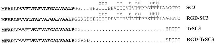
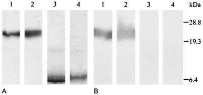
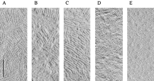
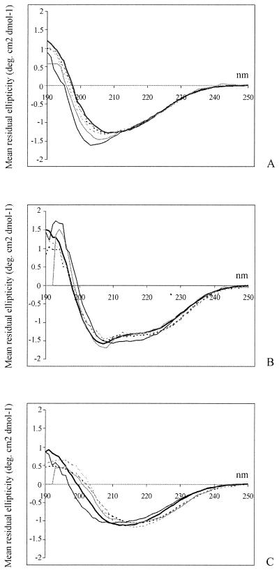
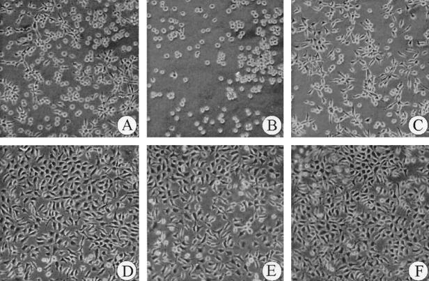
Hydrophobins are small (ca. 100 amino acids) secreted fungal proteins that are characterized by the presence of eight conserved cysteine residues and by a typical hydropathy pattern. Class I hydrophobins self-assemble at hydrophilic-hydrophobic interfaces into highly insoluble amphipathic membranes, thereby changing the nature of surfaces. Hydrophobic surfaces become hydrophilic, while hydrophilic surfaces become hydrophobic. To see whether surface properties of assembled hydrophobins can be changed, 25 N-terminal residues of the mature SC3 hydrophobin were deleted (TrSC3). In addition, the cell-binding domain of fibronectin (RGD) was fused to the N terminus of mature SC3 (RGD-SC3) and TrSC3 (RGD-TrSC3). Self-assembly and surface activity were not affected by these modifications. However, physiochemical properties at the hydrophilic side of the assembled hydrophobin did change. This was demonstrated by a change in wettability and by enhanced growth of fibroblasts on Teflon-coated with RGD-SC3, TrSC3, or RGD-TrSC3 compared to bare Teflon or Teflon coated with SC3. Thus, engineered hydrophobins can be used to functionalize surfaces.
Figures
References
-
- Ásgeirsdóttir, S. A., O. M. H. de Vries, and J. G. H. Wessels. 1998. Identification of three differentially expressed hydrophobins in Pleurotus ostreatus (oyster mushroom). Microbiology 144:2961-2969. - PubMed
-
- Beever, R. E., and G. P. Dempsey. 1978. Function of rodlets on the surface of fungal spores. Nature 272:608-610. - PubMed
-
- Bell-Pedersen, D., J. C. Dunlap, and J. J. Loros. 1992. The Neurospora circadian clock-controlled gene, ccg-2, is allelic to eas and encodes a fungal hydrophobin required for formation of the conidial rodlet layer. Genes Dev. 6:2382-2394. - PubMed
-
- Bowden, C. G., E. Smally, R. P. Guries, M. Hubbes, B. Temple, and P. A. Horgen. 1996. Lack of association between cerato-ulmin production and virulence in Ophiostoma ulmi. Mol. Plant-Microbe Interact. 9:556-564. - PubMed
-
- de Groot, P. W. J., P. J. Schaap, A. S. M. Sonneberg, J. Visser, and L. J. L. D. van Griensven. 1996. The Agaricus bisporus hypA gene encodes a hydrophobin and specifically accumulates in peel tissue of mushroom caps during fruit body development. J. Mol. Biol. 257:1008-1019. - PubMed
Publication types
MeSH terms
Substances
LinkOut - more resources
Full Text Sources
Other Literature Sources
