

doi: 10.1128/JB.184.6.1607-1616.2002.
Osmoregulation of dimer resolution at the plasmid pJHCMW1 mwr locus by Escherichia coli XerCD recombination
Affiliations
- PMID: 11872712
- PMCID: PMC134880
- DOI: 10.1128/JB.184.6.1607-1616.2002
Item in Clipboard
Osmoregulation of dimer resolution at the plasmid pJHCMW1 mwr locus by Escherichia coli XerCD recombination
J Bacteriol.
2002 Mar.
Abstract
Xer-mediated dimer resolution at the mwr site of plasmid pJHCMW1 is osmoregulated in Escherichia coli. Whereas under low-salt conditions, the site-specific recombination reaction is efficient, under high-salt conditions, it proceeds inefficiently. Regulation of dimer resolution is independent of H-NS and is mediated by changes in osmolarity rather than ionic effects. The low level of recombination at high salt concentrations can be overcome by high levels of PepA or by mutating the ARG box to a sequence closer to the E. coli ARG box consensus. The central region of the mwr core recombination site plays a role in regulation of site-specific recombination by the osmotic pressure of the medium.
Figures
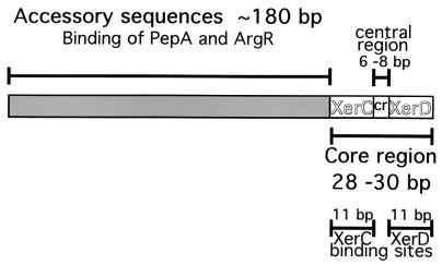
Schematic structure of plasmid Xer site-specific recombination sites. The sites contain a core recombination region that includes the XerC and XerD binding sites (11 bp each) and a central region (6 to 8 bp), as well as the accessory sequences (∼180 bp) with which ArgR and PepA interact (in the case of psi, ArcA and PepA). The diagram is not to scale. The hybrid sites utilized in this paper consist of accessory sequences (A) and core recombination regions (C) from different sites.
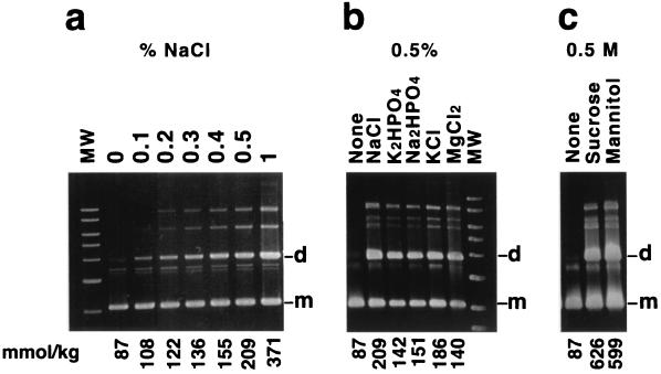
Resolution of pES dimers. Dimers were introduced by transformation into E. coli DS941. The cells were cultured in L medium with the addition of increasing NaCl concentrations (a); 0.5% NaCl, K2HPO4, Na2HPO4, KCl, and MgCl2 (b); or 0.5 M mannitol or sucrose (c). The osmolality values (millimoles per kilogram) are shown below each lane. The cultures were carried out in the presence of 100 μg of ampicillin per ml for 20 generations. Plasmid DNA was isolated and subjected to agarose gel electrophoresis. The slowly moving bands correspond to multimers (supercoiled and open circular). MW, linear molecular weight standards (10, 8, 6, 5, 4, 3, and 2 kb). d, dimer; m, monomer.
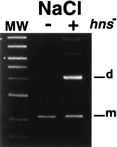
pES dimer resolution in an H-NS-deficient mutant. E. coli DS941 and E. coli DS9012 (H-NS deficient) harboring dimers of pES were cultured in L broth containing 0.5% (+) or no (−) NaCl, and plasmid DNA was purified and analyzed by gel agarose electrophoresis. The left lane shows linear DNA molecular weight (MW) standards (10, 8, 6, 5, 4, 3, 2, and 1.5 kb). d, dimer; m, monomer.
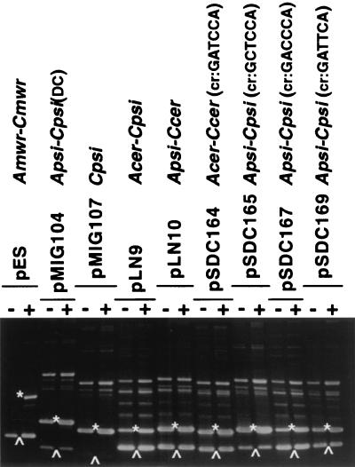
Xer site-specific recombination at several target sites. Dimers of pES and the different reporter plasmids were introduced by transformation into E. coli DS941. The cells were cultured in L medium containing 0.5% NaCl (+) or no NaCl (−). Plasmid DNA was isolated and subjected to agarose gel electrophoresis. The positions of unrecombined and recombined species are shown by asterisks and upward arrowheads, respectively.
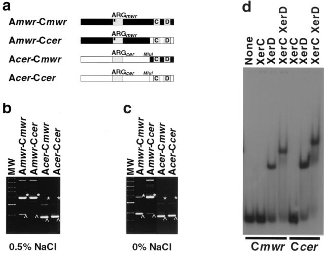
Resolution of dimers containing hybrid sites. (a) Recombination sites present in plasmids pKS492 (A cer-C cer), pES (A mwr-C mwr), pMET cm (A cer-C mwr), and pMET mc (A mwr-C cer). The bar in the ARG box in the mwr accessory sequences indicates that 1 nucleotide is different from the conserved consensus ARG box (ArgR binding site) sequence. (b and c) Dimers of pES, pMET mc, pMET cm, and pKS492 were introduced by transformation into E. coli DS941 and cultured in L medium containing 0.5% NaCl (b) or no NaCl (c). Plasmid DNA was isolated and subjected to agarose gel electrophoresis. Linear DNA molecular weight (MW) standards (10, 8, 6, 5, 4, 3, 2, and 1.5 kb) and the positions of dimer (asterisks) and monomer (arrowheads) species are shown. (d) In vitro protein-DNA binding. Oligonucleotides containing the mwr and cer core regions were end labeled and incubated in the presence of XerC, XerD, or both XerC and XerD. A control with no additions is shown. The products were separated by electrophoresis in 8% polyacrylamide gel.
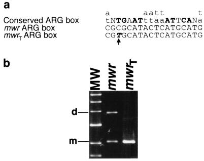
Mutagenesis of the mwr ARG box. (a) Nucleotide sequence of the mwr ARG box showing the substitution in mwrT and the consensus sequence of the ARG box according to Glansdorff (20). Boldface capital letters indicate the most important conserved nucleotides in the ARG box. (b) Dimers of pES (mwr) and pKD3 (mwrT) were introduced by transformation into E. coli DS941 and cultured in L broth containing 0.5% NaCl. Plasmid DNA was isolated and subjected to agarose gel electrophoresis. The left lane shows linear DNA molecular weight standards (6, 5, 4, 3, 2, and 1.5 kb). d, dimer; m, monomer.
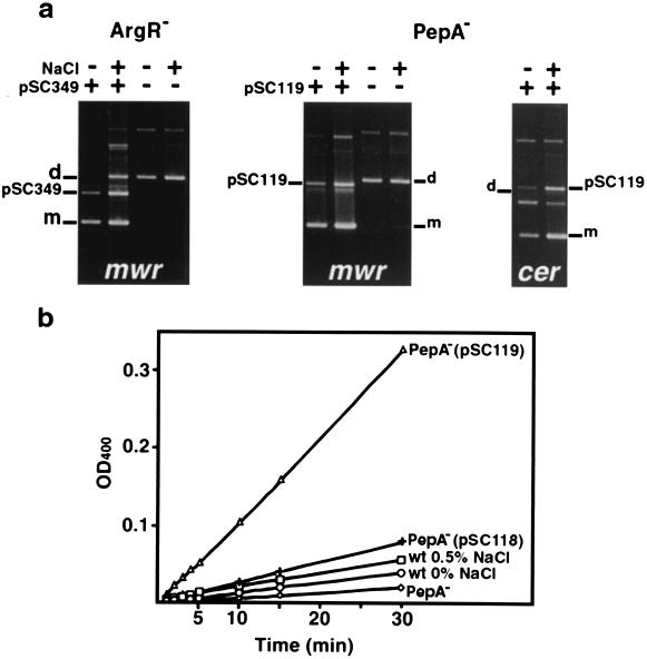
Complementation of ArgR− and PepA− derivatives. (a) E. coli DS956 (argR) and DS957 (pepA) were transformed with dimers of pES (A mwr-C mwr) or dimers of pKS492 (A cer-C cer) and pCS349 (argR gene fusion) or pCS119 (pepA gene fusion), respectively. These derivatives, as well as control E. coli DS956 and DS957 harboring only pES dimers, were cultured in L broth containing 0.5% NaCl (+) or no NaCl (−). Plasmid DNA was isolated and subjected to agarose gel electrophoresis. d, dimer; m, monomer. (b) Aminopeptidase A was partially purified, and its activity was determined as described before (26) for E. coli DS957 (◊), E. coli DS957(pSC118) (+), E. coli DS957(pSC119) (Δ), and E. coli DS941 cultured in L broth with no NaCl (○) or 0.5% NaCl (□).
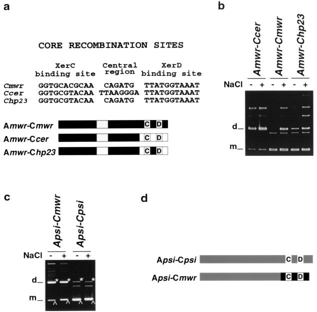
Resolution of dimers containing a modified core recombination site. (a) Comparison of the nucleotide sequence of core recombination sites of mwr, cer, and the mutant derivative called hp23 and diagram of the hybrid recombination site assayed. Gray boxes indicate the location of the XerC (C) and XerD (D) binding sites and the ARG box. (b and c) E. coli DS941 transformed with dimers harboring the indicated sites were cultured in L medium containing 0.5% NaCl (+) or no NaCl (−). Plasmid DNA was isolated and subjected to agarose gel electrophoresis. d, dimer; m, monomer.(d) Diagram representing A psi-C psi and the A psi-C mwr hybrid sites.
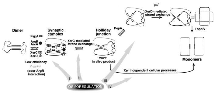
Osmoregulation of Xer recombination at mwr. The diagram shows the steps of the recombination process indicating proteins or features involved. For clarity, the proteins are shown only in the synaptic complex. The accessory proteins are PepA and ArgR (for cer and mwr) or PepA and ArcA (for psi). Bold lines represent accessory sequences. Small hexagons represent the C terminus of XerD activating XerC. The black circles represent the XerC tyrosine residue properly located to exchange the top strands. The Xer-mediated pathway of resolution of the Holliday junction at psi is shown at the top. The bottom shows the Xer-independent resolution pathway followed by cer (and probably by psi and mwr). For clarity, the supercoiling of the molecules is not shown. The ability to resolve through the Xer-dependent pathway is determined by the central region of the core recombination site (2). The gray arrows show possible levels of osmoregulation involving the central region.
Similar articles
-
Stability by multimer resolution of pJHCMW1 is due to the Tn1331 resolvase and not to the Escherichia coli Xer system.Microbiology (Reading). 2000 Mar;146 ( Pt 3):581-589. doi: 10.1099/00221287-146-3-581. Microbiology (Reading). 2000. PMID: 10746761
-
Differences in resolution of mwr-containing plasmid dimers mediated by the Klebsiella pneumoniae and Escherichia coli XerC recombinases: potential implications in dissemination of antibiotic resistance genes.J Bacteriol. 2006 Apr;188(8):2812-20. doi: 10.1128/JB.188.8.2812-2820.2006. J Bacteriol. 2006. PMID: 16585742 Free PMC article.
-
mwr Xer site-specific recombination is hypersensitive to DNA supercoiling.Nucleic Acids Res. 2009 Jun;37(11):3580-7. doi: 10.1093/nar/gkp208. Epub 2009 Apr 9. Nucleic Acids Res. 2009. PMID: 19359357 Free PMC article.
-
Timing, self-control and a sense of direction are the secrets of multicopy plasmid stability.Mol Microbiol. 1998 Sep;29(5):1137-45. doi: 10.1046/j.1365-2958.1998.01012.x. Mol Microbiol. 1998. PMID: 9767582 Review.
-
The topology of plasmid-monomerizing Xer site-specific recombination.Biochem Soc Trans. 2013 Apr;41(2):589-94. doi: 10.1042/BST20120340. Biochem Soc Trans. 2013. PMID: 23514159 Review.
Cited by
-
Functional Analysis of the Acinetobacter baumannii XerC and XerD Site-Specific Recombinases: Potential Role in Dissemination of Resistance Genes.Antibiotics (Basel). 2020 Jul 13;9(7):405. doi: 10.3390/antibiotics9070405. Antibiotics (Basel). 2020. PMID: 32668667 Free PMC article.
-
Small Klebsiella pneumoniae Plasmids: Neglected Contributors to Antibiotic Resistance.Front Microbiol. 2019 Sep 20;10:2182. doi: 10.3389/fmicb.2019.02182. eCollection 2019. Front Microbiol. 2019. PMID: 31616398 Free PMC article.
-
fpr, a deficient Xer recombination site from a Salmonella plasmid, fails to confer stability by dimer resolution: comparative studies with the pJHCMW1 mwr site.J Bacteriol. 2010 Feb;192(3):883-7. doi: 10.1128/JB.01082-09. Epub 2009 Dec 4. J Bacteriol. 2010. PMID: 19966005 Free PMC article.
-
Plasmid-Mediated Antibiotic Resistance and Virulence in Gram-negatives: the Klebsiella pneumoniae Paradigm.Microbiol Spectr. 2014;2(5):1-15. doi: 10.1128/microbiolspec.PLAS-0016-2013. Microbiol Spectr. 2014. PMID: 25705573 Free PMC article.
-
Complete nucleotide sequence of Klebsiella pneumoniae multiresistance plasmid pJHCMW1.Antimicrob Agents Chemother. 2002 Nov;46(11):3422-7. doi: 10.1128/AAC.46.11.3422-3427.2002. Antimicrob Agents Chemother. 2002. PMID: 12384346 Free PMC article.
References
-
- Arciszewska, L. K., R. A. Baker, B. Hallet, and D. J. Sherratt. 2000. Coordinated control of XerC and XerD catalytic activities during Holliday junction resolution. J. Mol. Biol. 299:391-403. - PubMed
-
- Blake, J. A., N. Ganguly, and D. J. Sherratt. 1997. DNA sequence of recombinase-binding sites can determine Xer site-specific recombination outcome. Mol. Microbiol. 23:387-398. - PubMed
Publication types
MeSH terms
Substances
Grants and funding
LinkOut - more resources
Full Text Sources
Other Literature Sources
Miscellaneous
