. 2002 Mar;17(3):493-501.
doi: 10.1359/jbmr.2002.17.3.493.

Noninvasive loading of the murine tibia: an in vivo model for the study of mechanotransduction

Affiliations

Noninvasive loading of the murine tibia: an in vivo model for the study of mechanotransduction

Ted S Gross et al. J Bone Miner Res. 2002 Mar.

Abstract

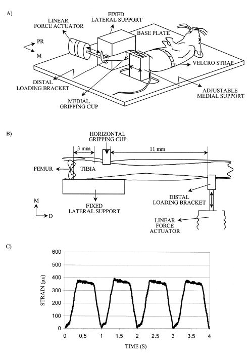
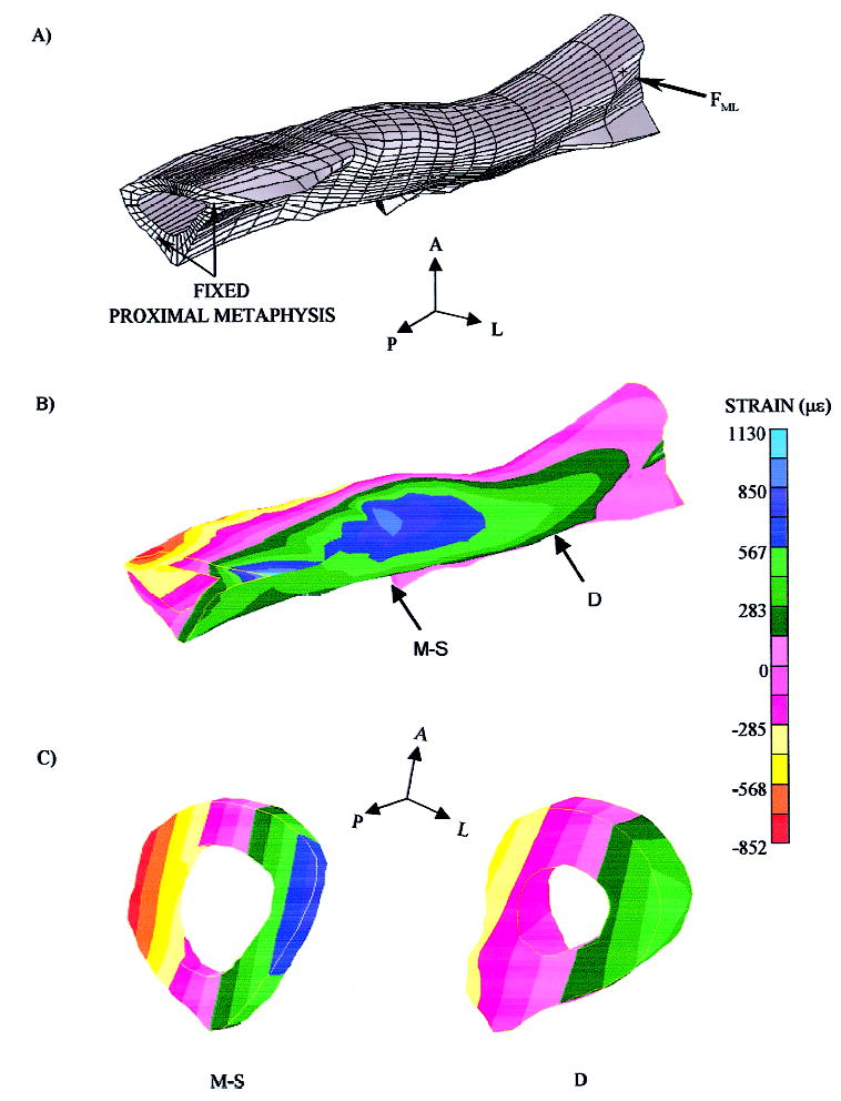
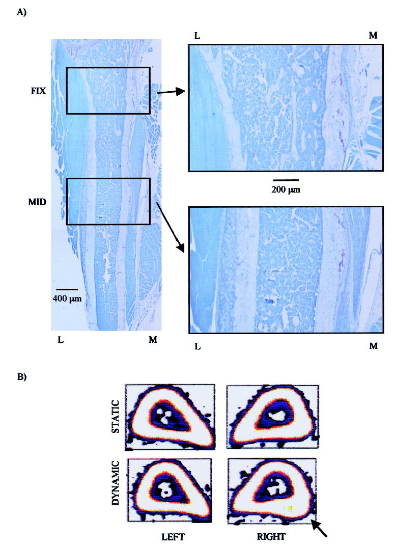
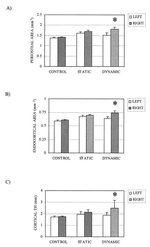
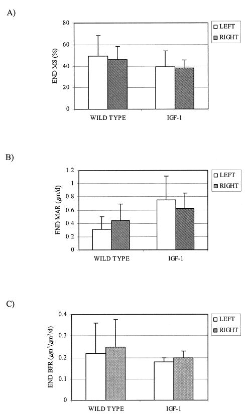
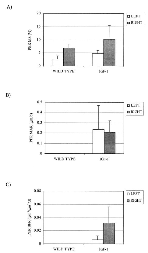
Transgenic and knockout mice present a unique opportunity to study mechanotransduction pathways in vivo, but the difficulty inherent with applying externally controlled loads to the small mouse skeleton has hampered this approach. We have developed a novel device that enables the noninvasive application of controlled mechanical loads to the murine tibia. Calibration of tissue strains induced by the device indicated that the normal strain environment was repeatable across loading bouts. Two in vivo studies were performed to show the usefulness of the device. Using C57Bl/6J mice, we found that dynamic but not static loading increased cortical bone area. This result is consistent with previous models of bone adaptation, and the lack of adaptation induced by static loading serves as a negative control for the device. In a preliminary study, transgenic mice selectively overexpressing insulin-like growth factor 1 (IGF-1) in osteoblasts underwent a low-magnitude loading regimen. Periosteal bone formation was elevated 5-fold in the IGF-1-overexpressing mice but was not elevated in wild-type littermates, showing the potential for synergism between mechanical loading and selected factors. Based on these data, we anticipate that the murine tibia-loading device will enhance assessment of mechanotransduction pathways in vivo and, as a result, has the potential to facilitate novel gene discovery and optimization of synergies between drug therapies and mechanical loading.


Conflict of interest statement

The authors have no conflict of interest.

Figures

FIG. 1
FIG. 1
(A) Schematic of the noninvasive murine loading device. Once anesthetized, the mouse is placed on its back and secured to the base plate. The proximal right tibia is secured at the metaphysis by a gripping cup attached to the adjustable medial support. (B) A computer controlled linear force actuator attached to a distal loading bracket applies small forces (0.3N is sufficient to induce physiological magnitude strains) to the distal tibial metaphysis (dorsal view). This design enables experimental control over the external loads applied to the tibia, while leaving the diaphysis free of contact. (C) Midshaft normal strains measured with a single element gage attached to the lateral surface indicate that the device induces a repeatable sawtooth waveform.
FIG. 2
FIG. 2
Full-field normal strain environment induced in the mouse tibia by the loading device. (A) An FE mesh was developed based on serial images of the tibia. As with the device, end loads (FML) were applied to the distal metaphysis, while the proximal metaphysis was fixed. (B and C) Normal strains were highly nonuniform in magnitude along the tibia diaphysis because of loading environment and morphology of the bone. For example, peak normal strains at the tibia midshaft (M-S) were approximately twice those observed at a (D) cross-section 3 mm distal to the midshaft.
FIG. 3
FIG. 3
Dynamic loading via the noninvasive murine loading device focally increases cortical bone area in C57Bl/6J mice. (A) A toluidine blue–stained frontal section of a tibia from a dynamically loaded mouse revealed no tissue reaction at the site of proximal fixation (FIX). At higher power, the unusual morphology on the medial (M) cortex was associated with the proximal tibial tuberosity. In contrast, higher power images of the midshaft (MID) revealed substantial new bone formation, predominantly on the lateral (L) cortex. This adaptive response was independent of the proximal fixation site. (B) Midshaft pQCT images (2.8 mm proximal to the tibia-fibular junction) from the left (nonloaded) and right (loaded) tibia of representative mice in the static loading groups revealed no macroscopic adaptation. pQCT images from the tibia exposed to dynamic loading showed cortical hypertrophy, with focal periosteal expansion readily evident (arrow).
FIG. 4
FIG. 4
Static histomorphometry revealed that increased cortical area in the dynamic loading group was achieved by a combination of significant expansion at both the mean (+SE) (A) periosteal and the (B) endocortical envelopes (*p < 0.05). (C) Periosteal expansion outweighed the endocortical expansion, as mean cortical thickness was elevated over 30% in the dynamic loading group (*p < 0.05).
FIG. 5
FIG. 5
Low-magnitude loading did not alter endocortical osteoblastic activity in wild-type or IGF-1– overexpressing mice (n = 3 per group). No loading-related differences were observed in mean (+SE) endocortical (A) MS, (B) MAR, or (C) BFR. Endocortical BFR was nearly an order of magnitude greater than that observed on the periosteal surface.
FIG. 6
FIG. 6
Low-magnitude loading synergistically enhances periosteal osteoblastic activity in IGF-1– overexpressing mice (n = 3 per group). (A) Mechanical loading doubled the mean (+SE) percent MS in both wild-type and IGF-1 mice. However, dLSs were evident only in the loaded tibia of IGF-1 mice, and, as a result, (B and C) MAR and BFR in the contralateral left tibia of wild-type littermates and IGF-1 mice were negligible. Likewise, low-magnitude loading of the right tibia of wild-type littermates did not induce periosteal bone formation. However, the same loading regimen elevated periosteal bone formation 5-fold in the IGF-1 mice.

Similar articles

Cited by

References

    1. Riggs BL, Melton LJ. Involutional osteoporosis. N Engl J Med. 1986;314:1676–1686. - PubMed
    1. Cummings SR, Nevitt MC, Browner WS, Stone K, Fox KM, Ensrud KE, Cauley J, Black D, Vogt TM. Risk factors for hip fracture in white women. Study of Osteoporotic Fractures Research Group. N Engl J Med. 1995;332:767–773. - PubMed
    1. Liberman UA, Weiss SR, Broll J, Minne HW, Quan H, Bell NH, Rodriquez-Portales J, Downs RW, Dequeker J, Favus M. Effect of oral alendronate on bone mineral density and the incidence of fractures in postmenopausal osteoporosis. The Alendronate Phase III osteoporosis treatment study group. N Engl J Med. 1995;333:1437–1443. - PubMed
    1. NIAMS 2000 Bone biology and bone diseases research: Strategic plan (FY 2000 –2004). www.nih.gov/niams/strategicplan/bone.htm
    1. Rubin CT, Lanyon LE. Regulation of bone mass by mechanical strain magnitude. Calcif Tissue Int. 1985;37:411–417. - PubMed

Publication types

Substances