

Immunocytochemical method for early laboratory diagnosis of tuberculous meningitis
- PMID: 11874875
- PMCID: PMC119925
- DOI: 10.1128/cdli.9.2.344-347.2002
Immunocytochemical method for early laboratory diagnosis of tuberculous meningitis
Abstract
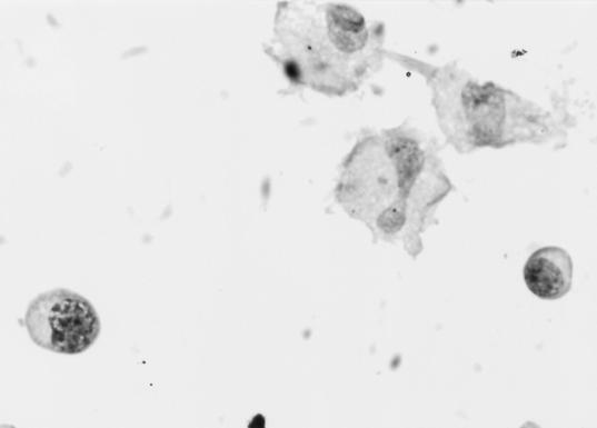
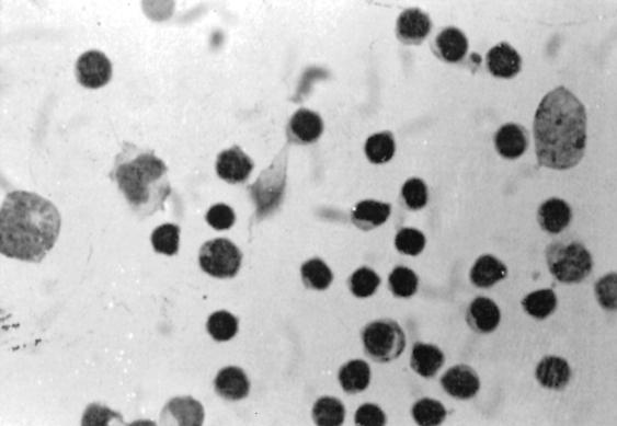
A simple immunocytochemical method was standardized for the direct demonstration of mycobacterial antigen in cerebrospinal fluid (CSF) specimens of patients with tuberculous meningitis (TBM). CSF-cytospin smears were prepared from 22 patients with a clinical diagnosis of TBM and also from an equal number of patients with nontuberculous neurological diseases (disease control). Immunocytological demonstration of mycobacterial antigens in the cytoplasm of monocytoid cells was attempted, by using rabbit immunoglobulin G to Mycobacterium tuberculosis as the primary antibody. Of the 22 CSF-cytospin smears from TBM patients, 16 showed positive immunostaining, while all of the CSF-cytospin smears from the disease control showed negative immunostaining for mycobacterial antigen. The technical aspects of this immunocytological method for the demonstration of mycobacterial antigens are simple, rapid, and reproducible, as well as specific, and therefore can be applied for the early diagnosis of TBM, particularly in patients in whom bacteriological methods did not demonstrate the presence of M. tuberculosis in the CSF.
Figures
References
-
- Annamma, M., V. V. Radhakrishnan, and S. Sehgal. 1990. Diagnosis of tuberculous meningitis by enzyme-linked immunosorbant assays to detect mycobacterial antigen and antibody in cerebrospinal fluid of patients with tuberculous meningitis. Med. Microbiol. Immunol. 179:281-288. - PubMed
-
- Bal, V. R., J. Kamat, J. Kamath, and P. Kadoth. 1983. Enzyme linked immunosorbent assay for mycobacterial antigens. Ind. J. Med. Res. 78:477-483. - PubMed
-
- Kox, L. F. F., S. Kuijper, and A. H. J. Kolk. 1995. Early diagnosis of tuberculous meningitis by polymerase chain reaction. Neurology 45:2228-2232. - PubMed
-
- Krambovitis, E., M. B. McIllmurray, P. E. Lock, W. Hendrickse, and H. Holzel. 1984. Rapid diagnosis of tuberculous meningitis by latex particle agglutination. Lancet ii:1229-1231. - PubMed
Publication types
MeSH terms
Substances
LinkOut - more resources
Full Text Sources
