. 2002 Mar;9(2):461-9.
doi: 10.1128/cdli.9.2.461-469.2002.

Detection of Cryptococcus neoformans DNA in tissue samples by nested and real-time PCR assays

Affiliations

Detection of Cryptococcus neoformans DNA in tissue samples by nested and real-time PCR assays

Ralf Bialek et al. Clin Diagn Lab Immunol. 2002 Mar.

Abstract

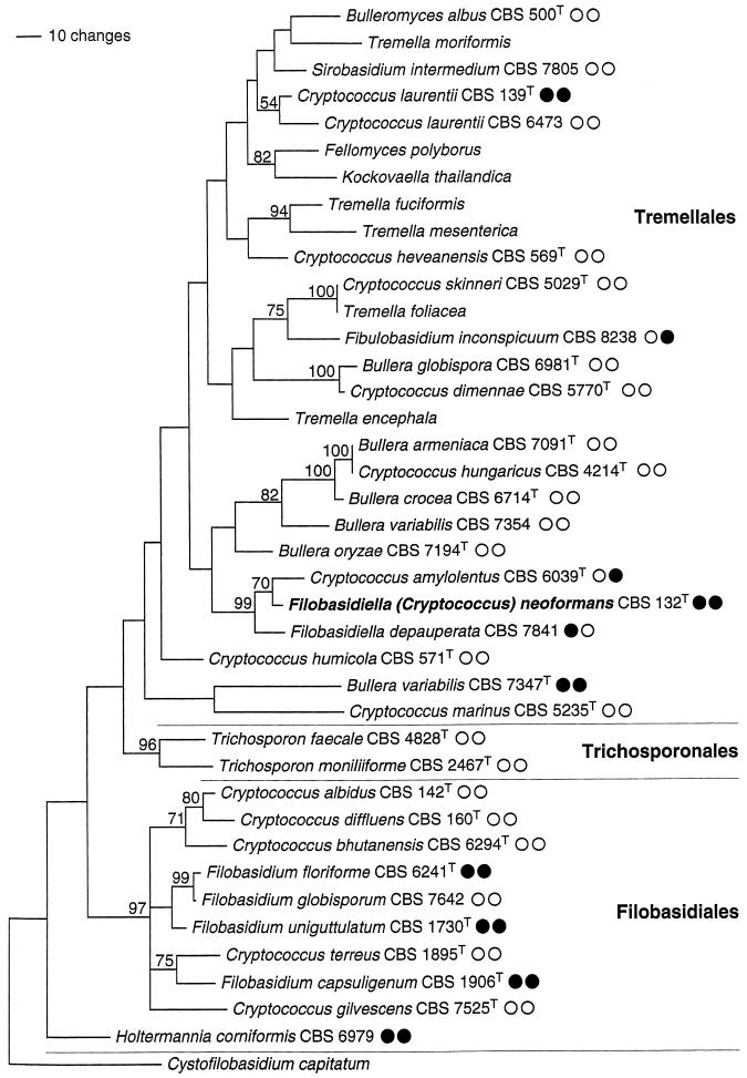
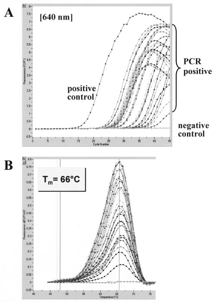
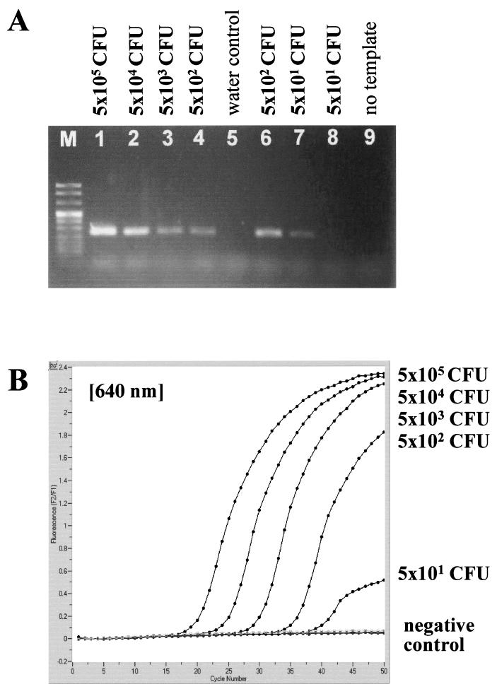
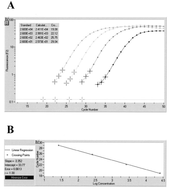
Two PCR protocols targeting the 18S rRNA gene of Cryptococcus neoformans were established, compared, and evaluated in murine cryptococcal meningitis. One protocol was designed as a nested PCR to be performed in conventional block thermal cyclers. The other protocol was designed as a quantitative single-round PCR adapted to LightCycler technology. One hundred brain homogenates and dilutions originating from 20 ICR mice treated with different azoles were examined. A fungal burden of 3 x 10(1) to 2.9 x 10(4) CFU per mg of brain tissue was determined by quantitative culture. Specific PCR products were amplified by the conventional and the LightCycler methods in 86 and 87 samples, respectively, with products identified by DNA sequencing and real-time fluorescence detection. An analytical sensitivity of 1 CFU of C. neoformans per mg of brain tissue and less than 10 CFU per volume used for extraction was observed for both PCR protocols, while homogenates of 70 organs from mice infected with other fungi were PCR negative. Specificity testing was performed with genomic DNA from 31 hymenomycetous fungal species and from the ustilaginomycetous yeast Malassezia furfur, which are phylogenetically related to C. neoformans. Twenty-four strains, including species of human skin flora like M. furfur and Trichosporon spp., were PCR negative. Amplification was observed with Cryptococcus amylolentus, Filobasidiella depauperata, Cryptococcus laurentii, and five species unrelated to clinical specimens. LightCycler PCR products from F. depauperata and Trichosporon faecale could be clearly discriminated by melting curve analysis. The sensitive and specific nested PCR assay as well as the rapid and quantitative LightCycler PCR assay might be useful for the diagnosis and monitoring of human cryptococcal infections.


Figures

FIG. 1.
FIG. 1.
Estimate of phylogenetic relationships in the Tremellomycetidae. Maximum parsimony analysis of an alignment of nuclear DNA sequences coding for the D1/D2 region of the ribosomal large subunit. Strict consensus of the three most parsimonious trees found in 1,000 rounds of heuristic search. Numbers on branches are bootstrap values from 1,000 replicates. The topology was rooted with Cystofilobasidium capitatum. Strain numbers are given for the species used in the PCR experiments of this study. CBS, Centraalbureau voor Schimmelcultures; T superscript, type strain; open and solid circles, negative and positive reaction results, respectively, of nested-PCR and LightCycler hybridization experiments.
FIG. 2.
FIG. 2.
Evaluation of the LightCycler PCR assay with brain tissue sections of C. neoformans-infected mice. A representative set of 28 samples with various C. neoformans concentrations was tested for the presence of C. neoformans 18S rDNA. Amplicon curves representing C. neoformans PCR-positive isolates are indicated by braces (A), and the corresponding results in melting curve analysis are depicted (B).
FIG. 3.
FIG. 3.
Analytical sensitivity of nested PCR (A) and LightCycler PCR (B) for the C. neoformans small-subunit rRNA gene (18S rDNA), determined with serial dilutions of a cultured C. neoformans strain. Detection limits in CFU per milliliter, determined by standard plating procedure, are given at the corresponding lanes in agarose gel electrophoresis or next to the LightCycler amplicon curves.
FIG. 4.
FIG. 4.
Quantitative analysis of LightCycler results obtained on a representative set of four tissue sections containing 2.6 × 101 to 2.6 × 10⁴ CFU of C. neoformans per processed sample volume. Based on the slope of the original amplification curves during the log-linear phase determined by three data points, artificial crossing points with the noise band (horizontal line) were determined by the LightCycler software (A). The standard curve represents the linear regression line through the data points on a plot of crossing points (threshold cycle) versus logarithm of standard sample concentration (B). Slope, y-intercept, mean squared error, and regression coefficient of the standard curve are given.

References

    1. Bialek, R., A. Ibricevic, C. Aepinus, L. K. Najvar, A. W. Fothergill, J. Knobloch, and J. R. Graybill. 2000. Detection of Paracoccidioides brasiliensis in tissue samples by a nested PCR assay. J. Clin. Microbiol. 38:2940-2942. - PMC - PubMed
    1. Bialek, R., J. Fischer, A. Feucht, L. K. Najvar, K. Dietz, J. Knobloch, and J. R. Graybill. 2001. Diagnosis and monitoring of murine histoplasmosis by a nested PCR assay. J. Clin. Microbiol. 39:1506-1509. - PMC - PubMed
    1. Blevins, L. B., J. Fenn, H. Segal, P. Newcomb-Gayman, and K. C. Carroll. 1995. False-positive cryptococcal antigen latex agglutination caused by disinfectants and soaps. J. Clin. Microbiol. 33:1674-1675. - PMC - PubMed
    1. Cheng, M.-F., C. C. Chiou, Y.-C. Liu, H.-Z. Wang, and K.-S. Hsieh. 2001. Cryptococcus laurentii fungemia in a premature neonate. J. Clin. Microbiol. 39:1608-1611. - PMC - PubMed
    1. Evertsson, U., H. J. Monstein, and A. G. Johansson. 2000. Detection and identification of fungi in blood using broad-range 28S rDNA PCR amplification and species-specific hybridisation. APMIS 108:385-392. - PubMed

LinkOut - more resources